Annotation summary for ref(2)P in insitu_ovaries_public
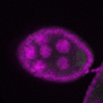
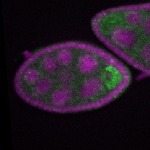
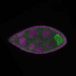
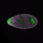
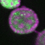
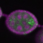
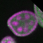
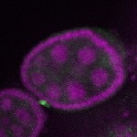
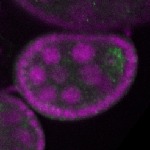
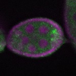
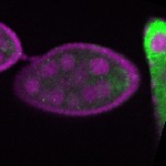
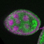
Legend: RNA signal: green, DNA staining: magenta
| Probe source: | GH06306-dg (Flybase link CG10360 , FlyFish insitu , BDGP insitu new) | |
| stage | images | Annotation |
| stage1 | 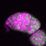 s s
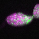 s s | |
| stage2-7 | 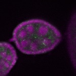 s s
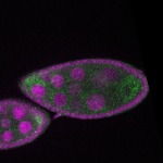 s s
 s s
 s s
 s s
 s s
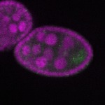 s s
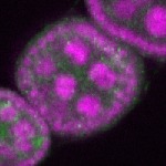 s s
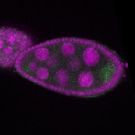 s s
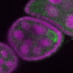 s s
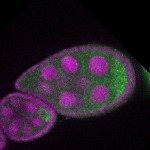 s s
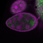 s s
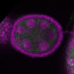 s s
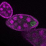 s s
 s s
 s s
 s s
 s s
 s s
 s s
 s s
 s s
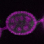 s s
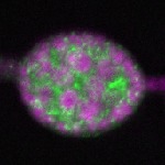 s s
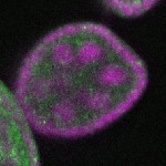 s s
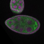 s s
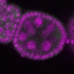 s s
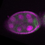 s s
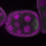 s s | enrichment_ubiquitous oocyte |
| stage8 | 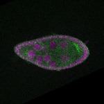 s s
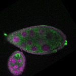 s s
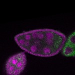 s s
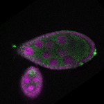 s s
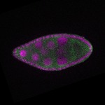 s s
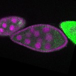 s s
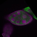 s s | ubiquitous signal |
| stage9 | 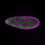 s s
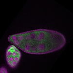 s s
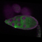 s s | cortical enrichment oocyte ubiquitous signal |
| stage10 | 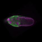 s s
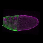 s s
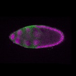 s s
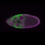 s s
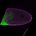 s s
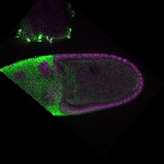 s s
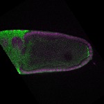 s s
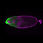 s s
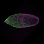 s s | cortical enrichment oocyte |
| Probe source: | RE24064 (Flybase link CG10360 , FlyFish insitu , BDGP insitu new) | |
| stage | images | Annotation |
| stage1 | 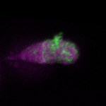 s s
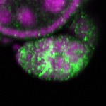 s s
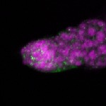 s s
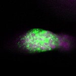 s s | |
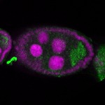
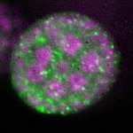
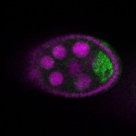
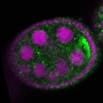
| stage2-7 | 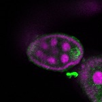 s s
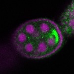 s s
 s s
 s s
 s s
 s s
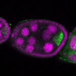 s s
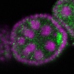 s s
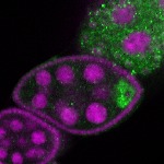 s s
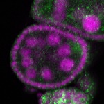 s s
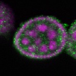 s s | enrichment_ubiquitous oocyte |
| stage8 | 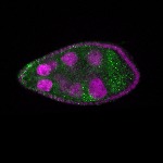 s s
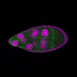 s s
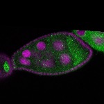 s s | ubiquitous signal |
| stage9 | 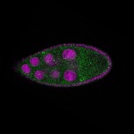 s s
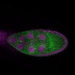 s s
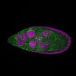 s s
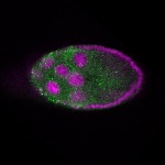 s s | ubiquitous signal |
| stage10 | 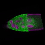 s s
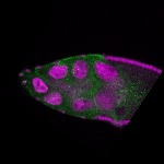 s s
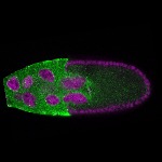 s s
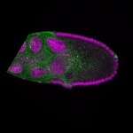 s s | ubiquitous signal |
| Probe source: | GH06306 (Flybase link CG10360 , FlyFish insitu , BDGP insitu new) | |
| No images or annotation terms | ||
Annotation summary for ref(2)P in insitu_fish
Legend: RNA signal: green, DNA staining: magenta
| Probe source: | GH06306 (Flybase link CG10360 , FlyFish insitu , BDGP insitu new) | |
| stage | images | Annotation |
| stage1-3 | 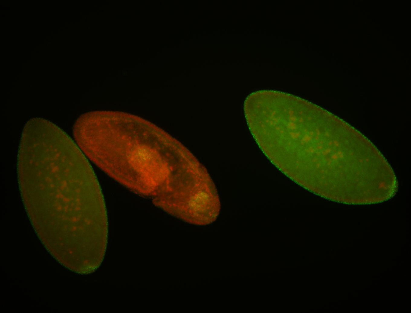

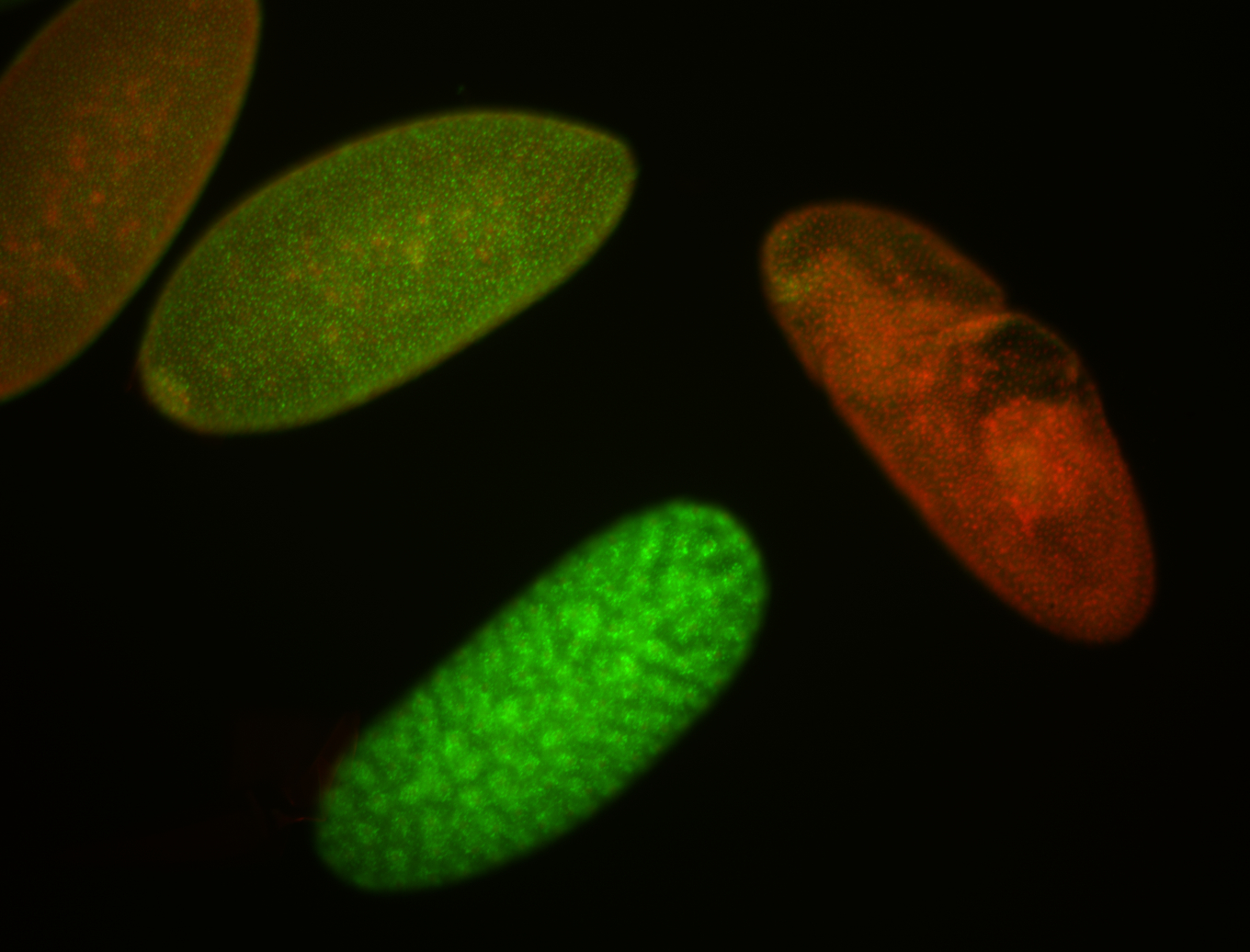
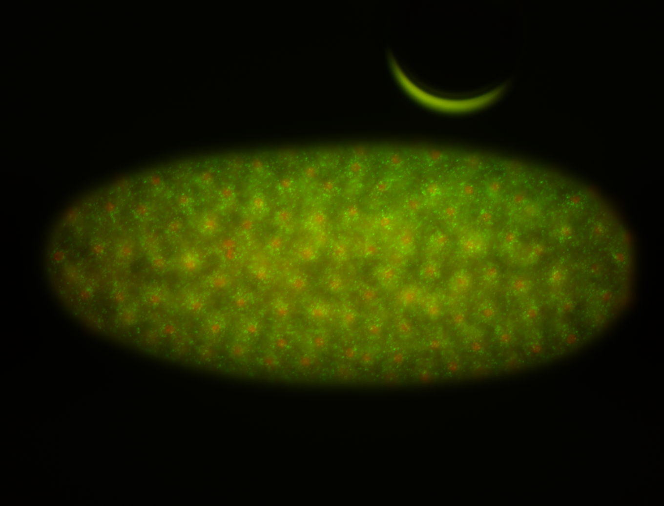
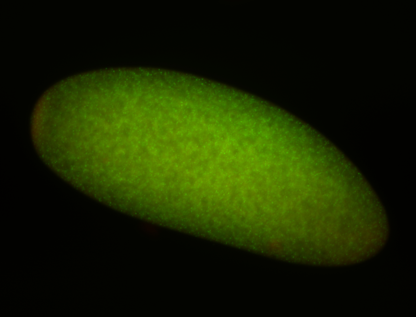
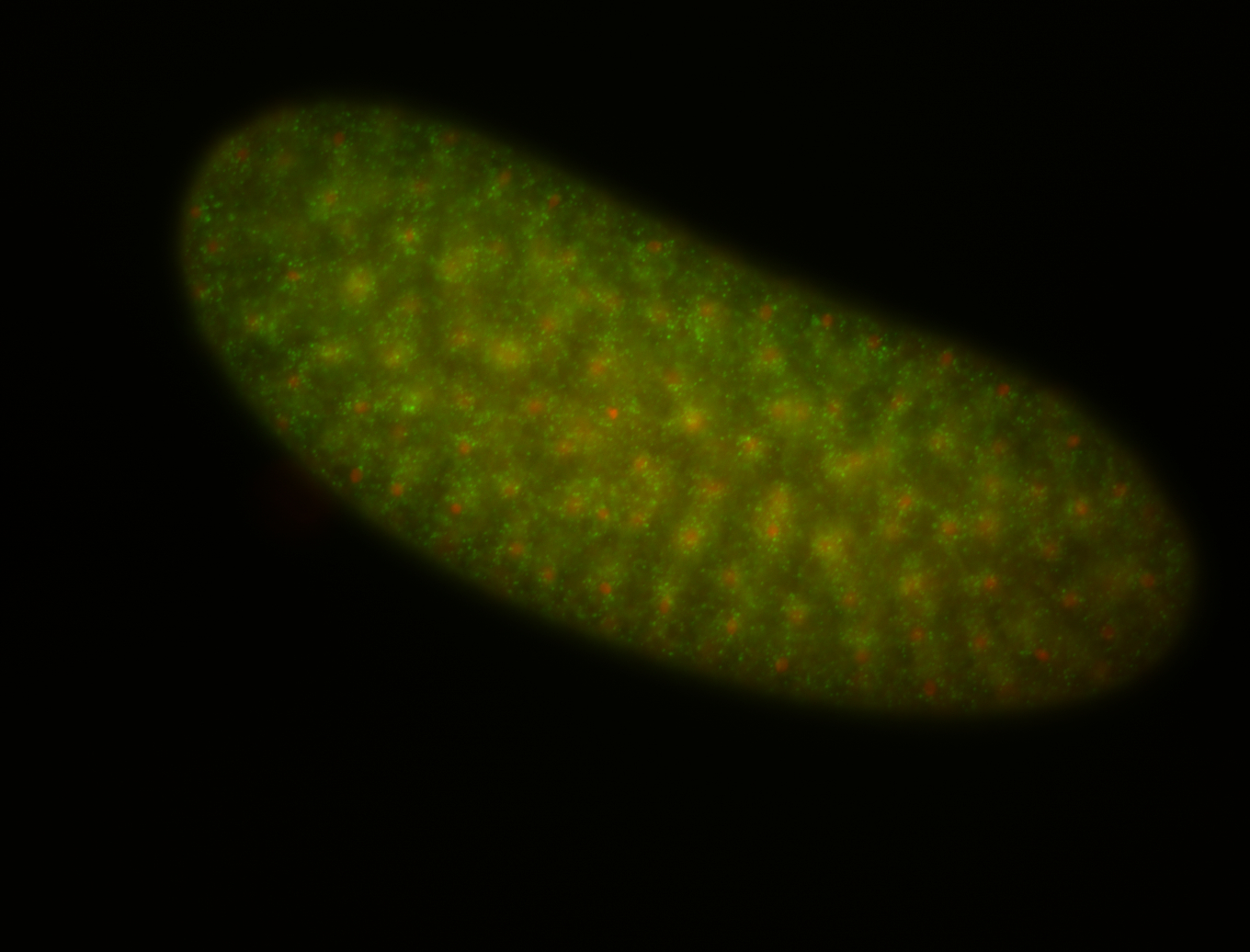
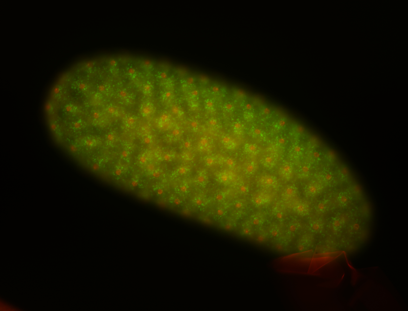
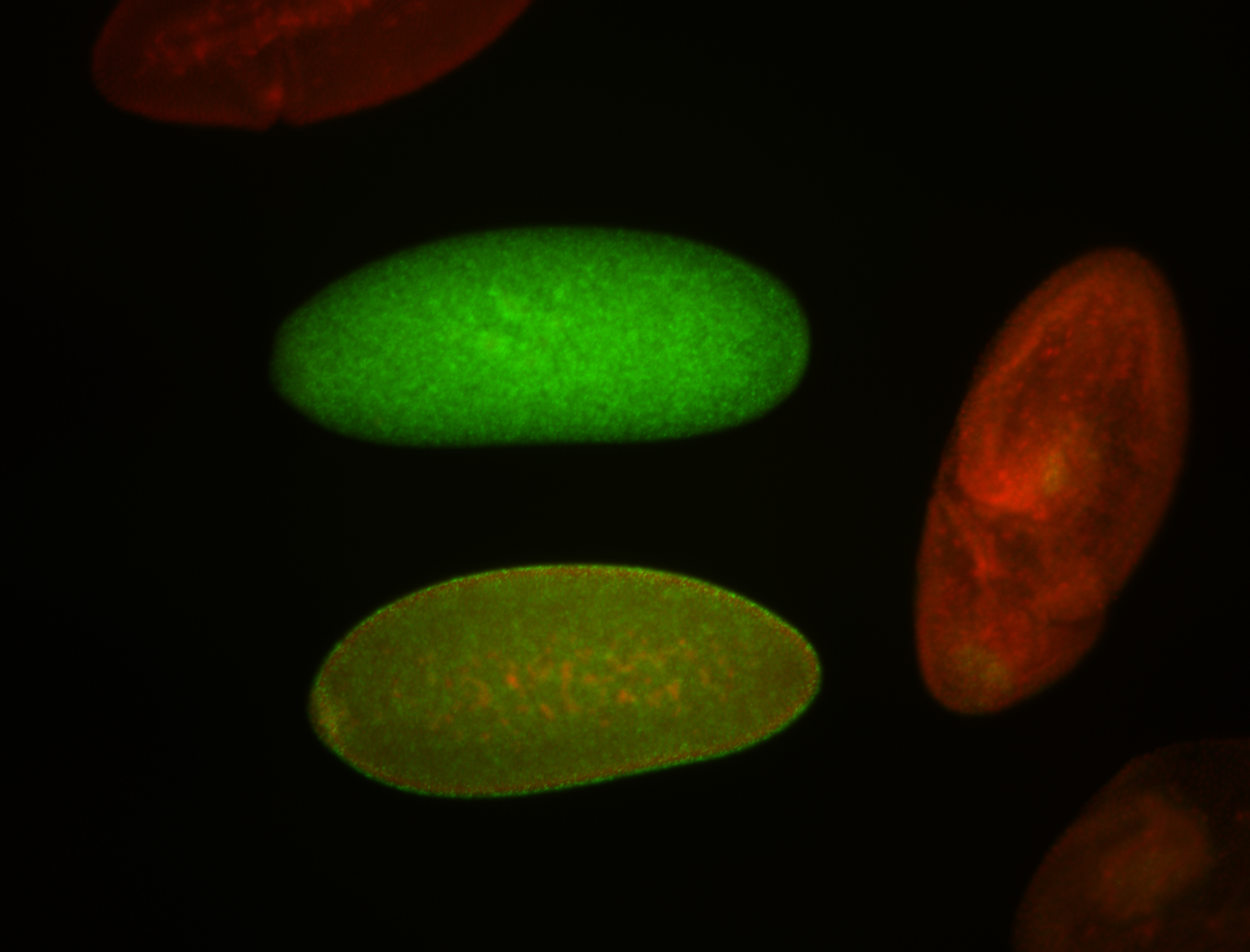
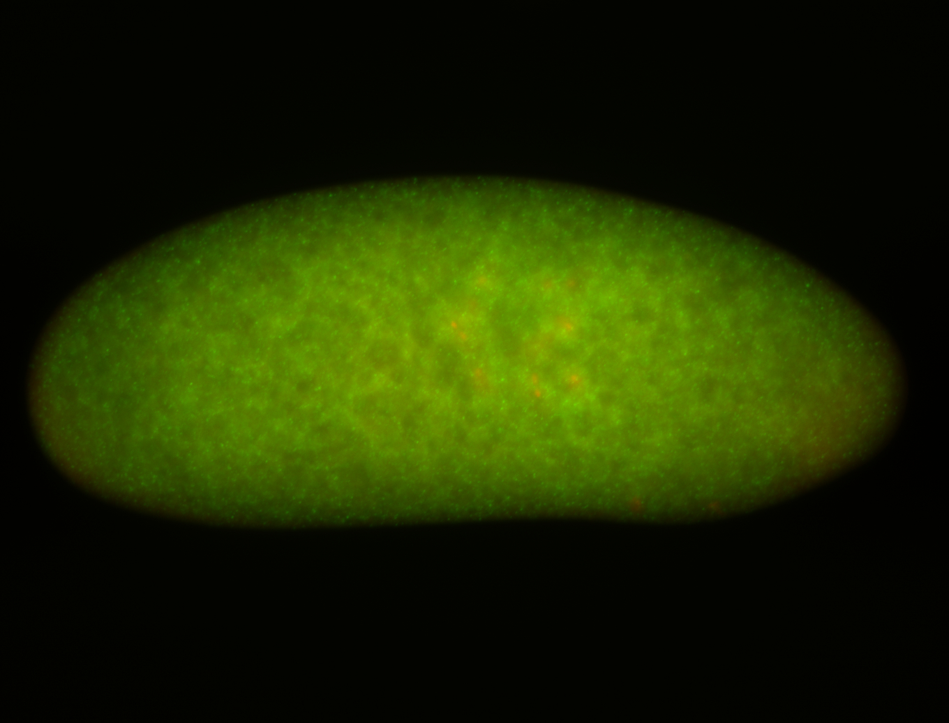
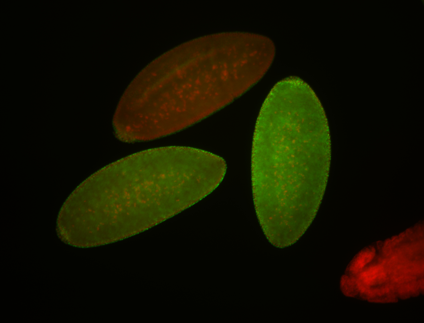 | Stage 1-3 Subcellular localization patterns Perinuclear localization Cell division apparatus Expressed Localized Perinuclear around all nuclei Embryonic localization patterns Nuclei associated localization Strong expression (1-20ms exp) Cytoplasmic foci Annotated by Ronit Maternal |
| stage4-5 | 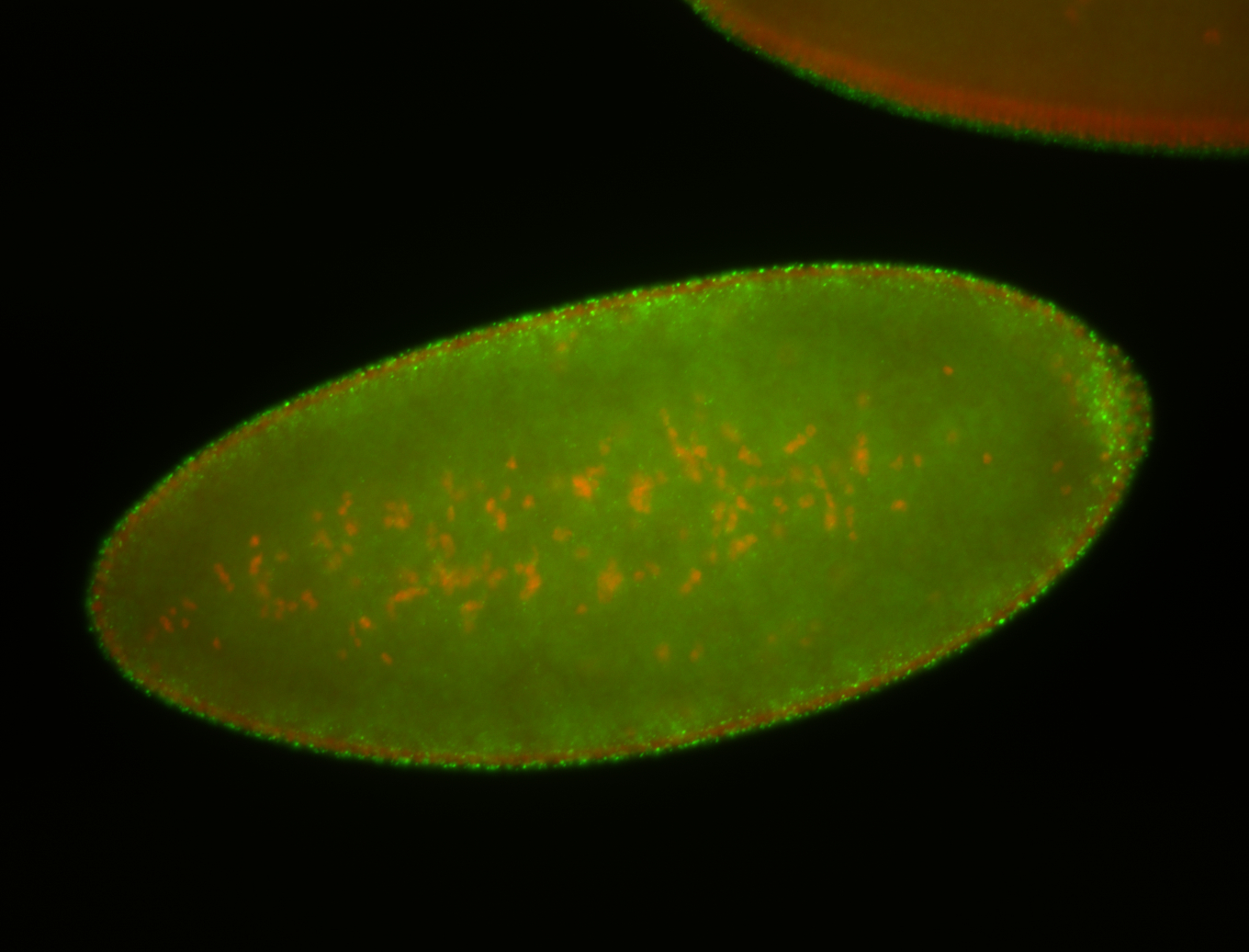
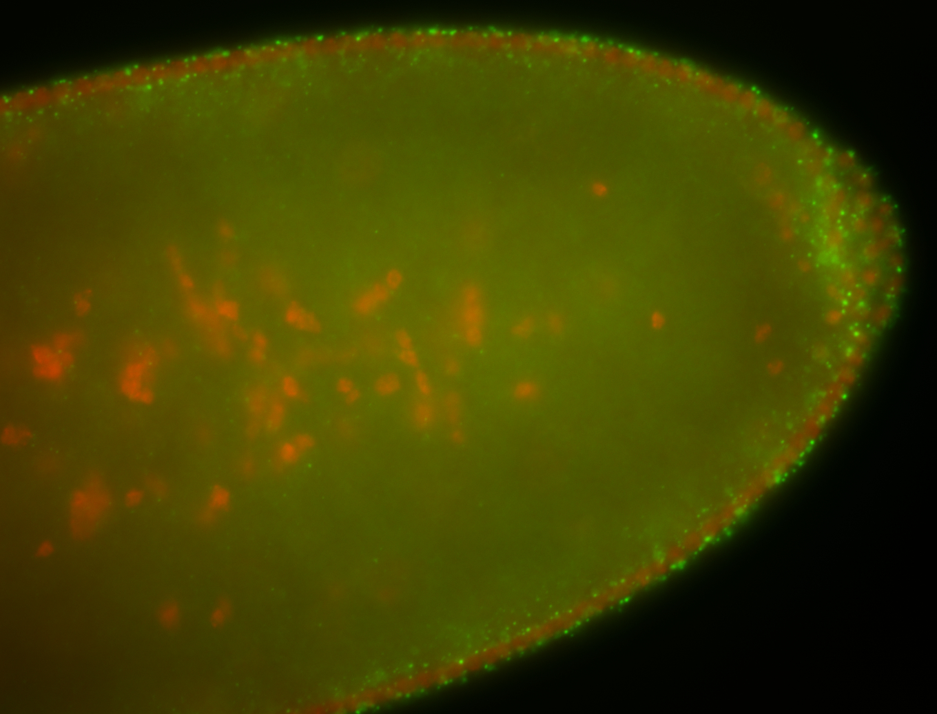
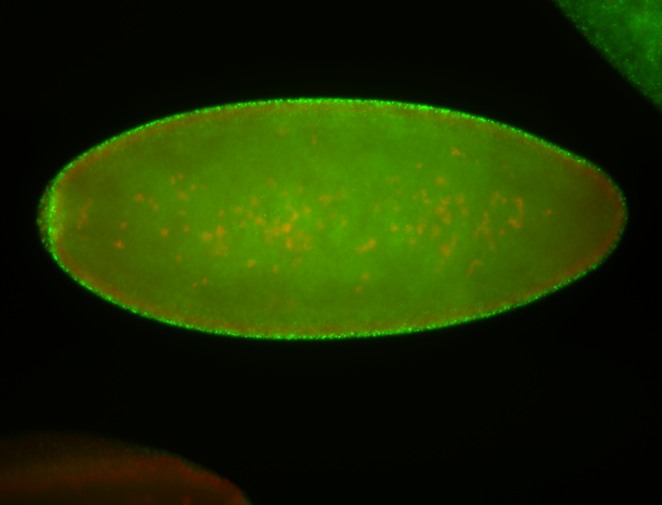
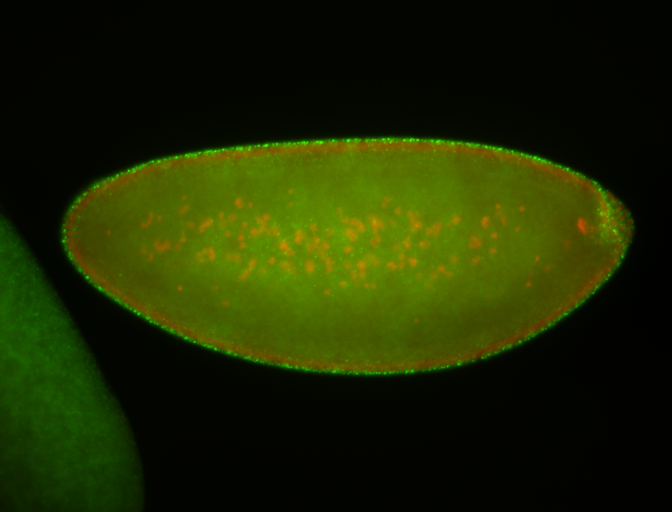
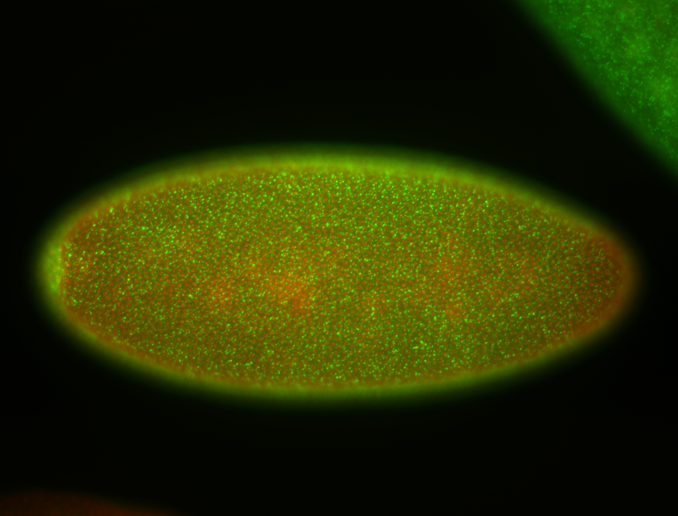


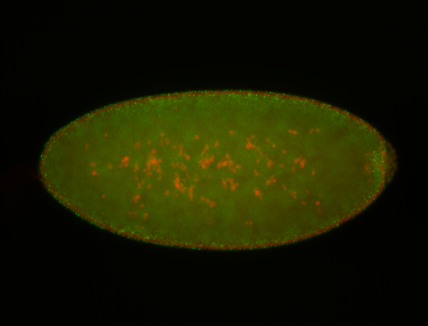
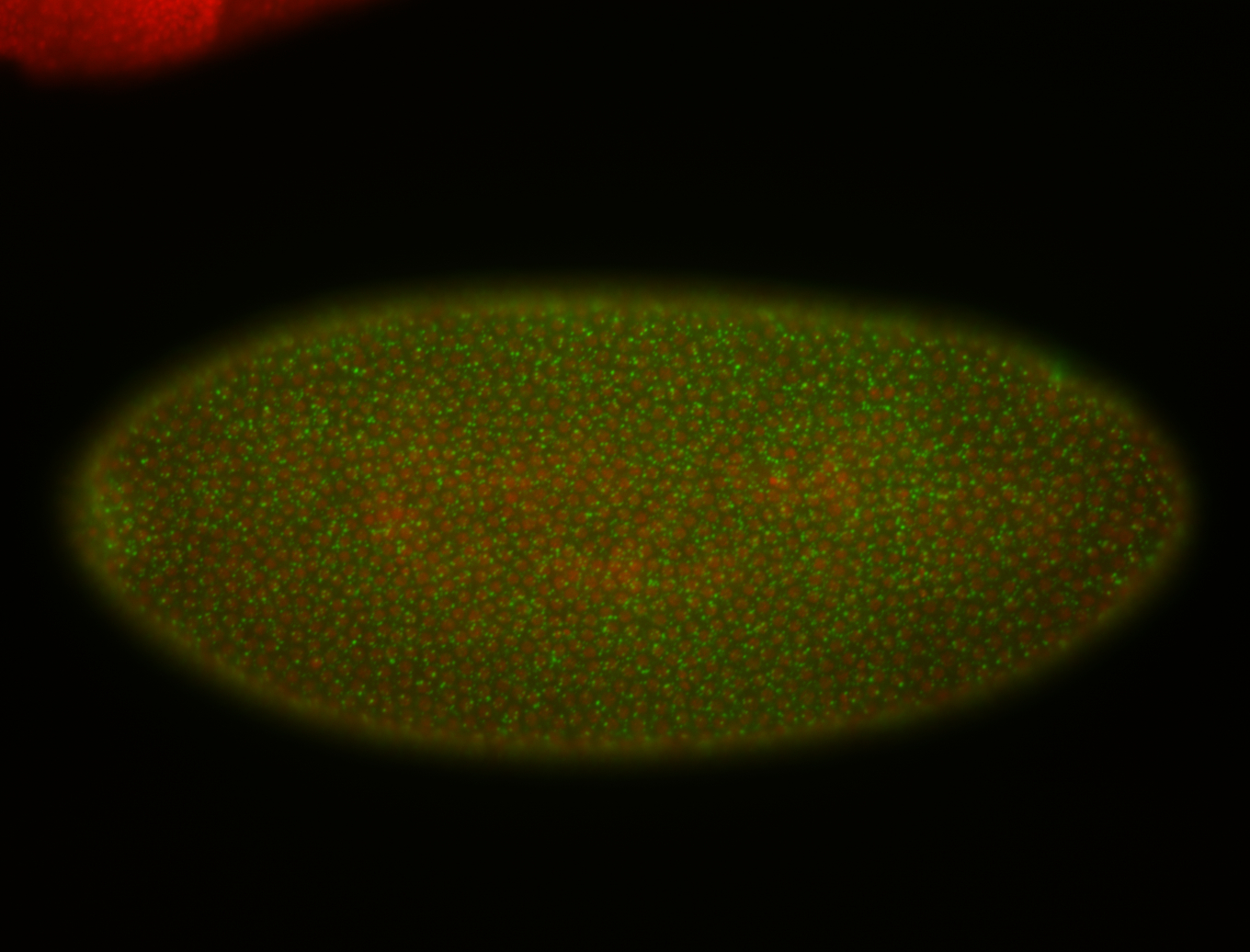
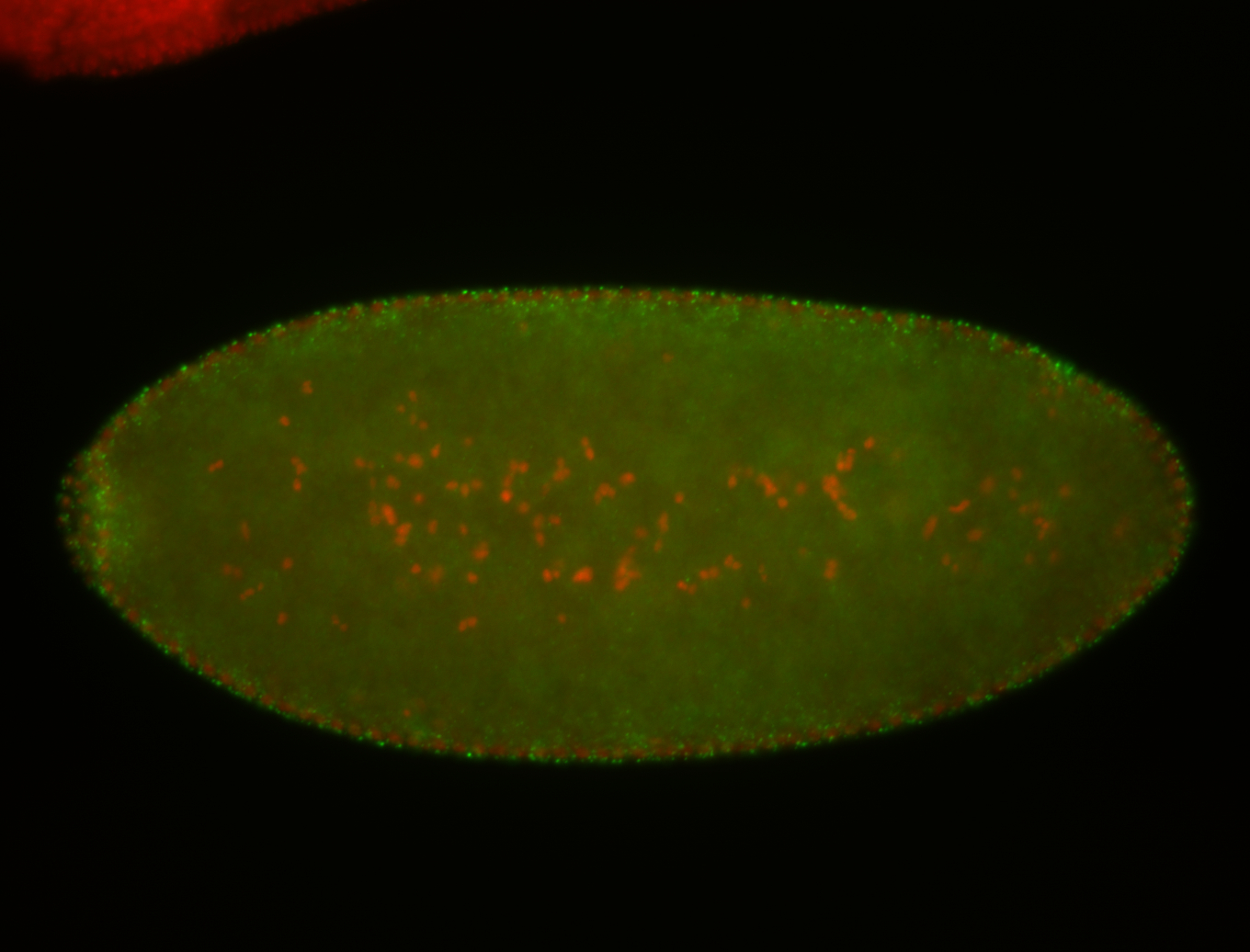
 | Cytoplasmic foci Degraded completely stage 5 Degraded partially stage 5 Pole cell localization Apical enrichment Discrete apical foci Stage 4-5 Embryonic localization patterns Localized Expressed Degraded Apical localization Pole cell enrichment Subcellular localization patterns Maternal |
| stage6-7 | 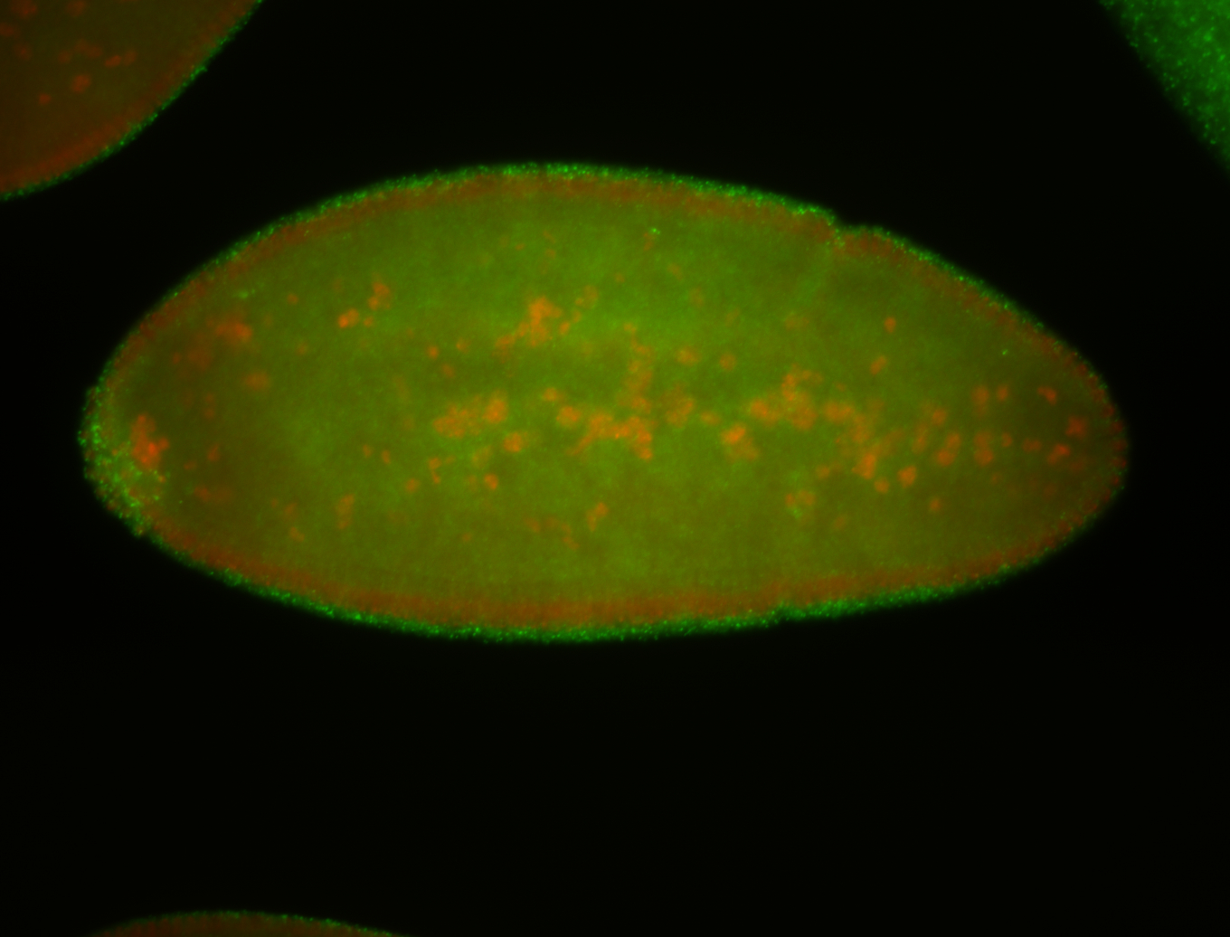
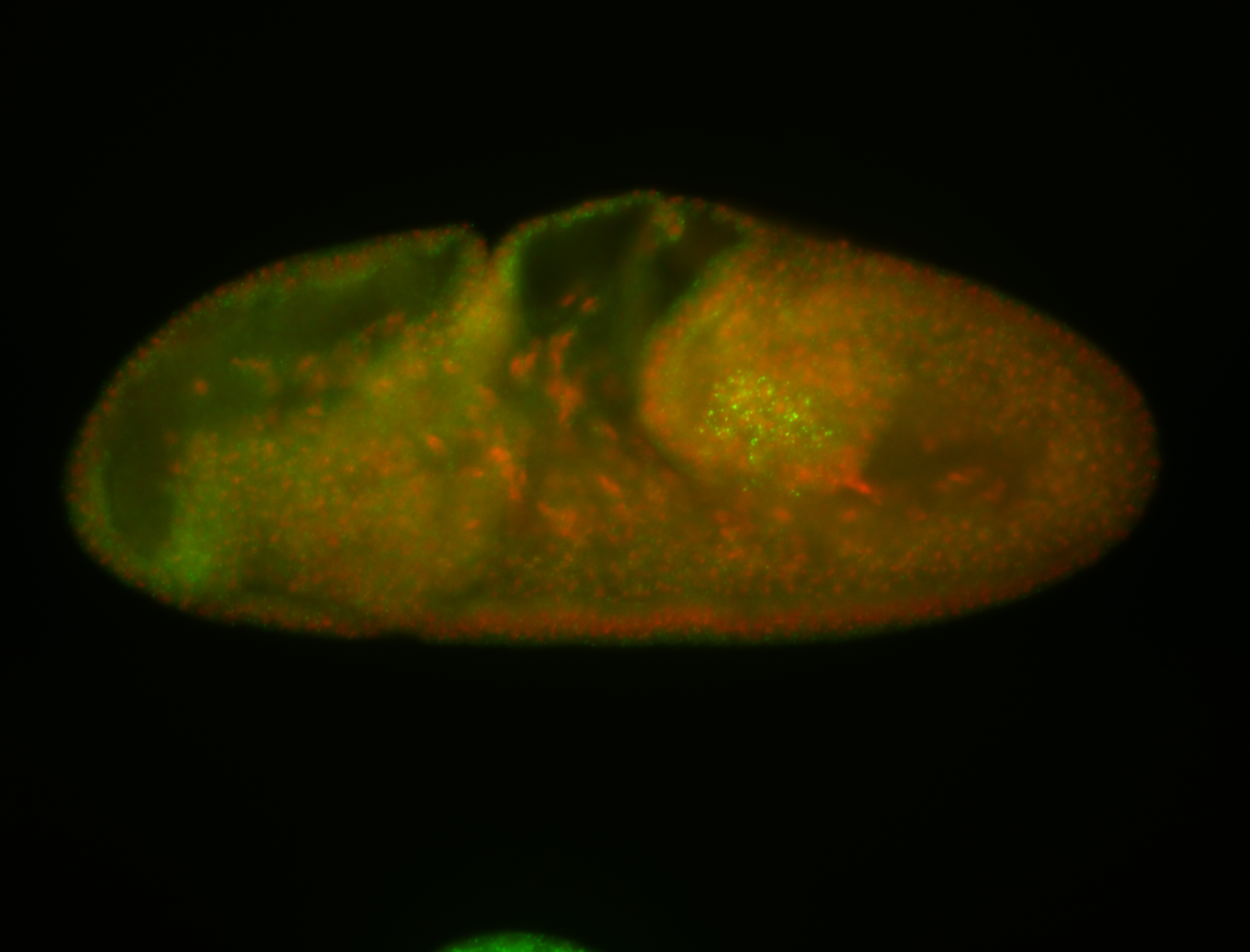
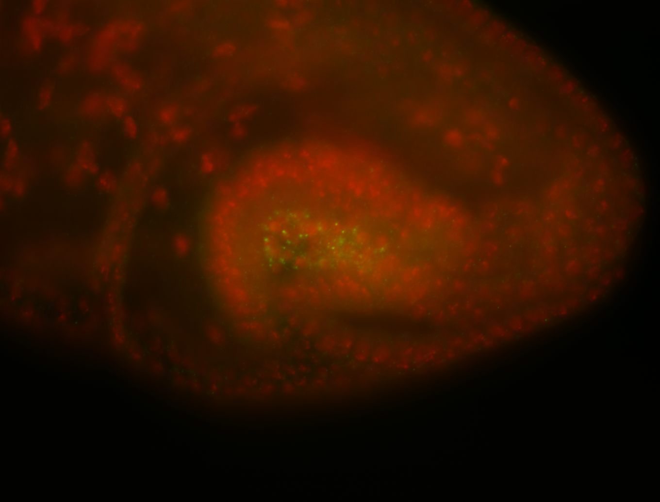
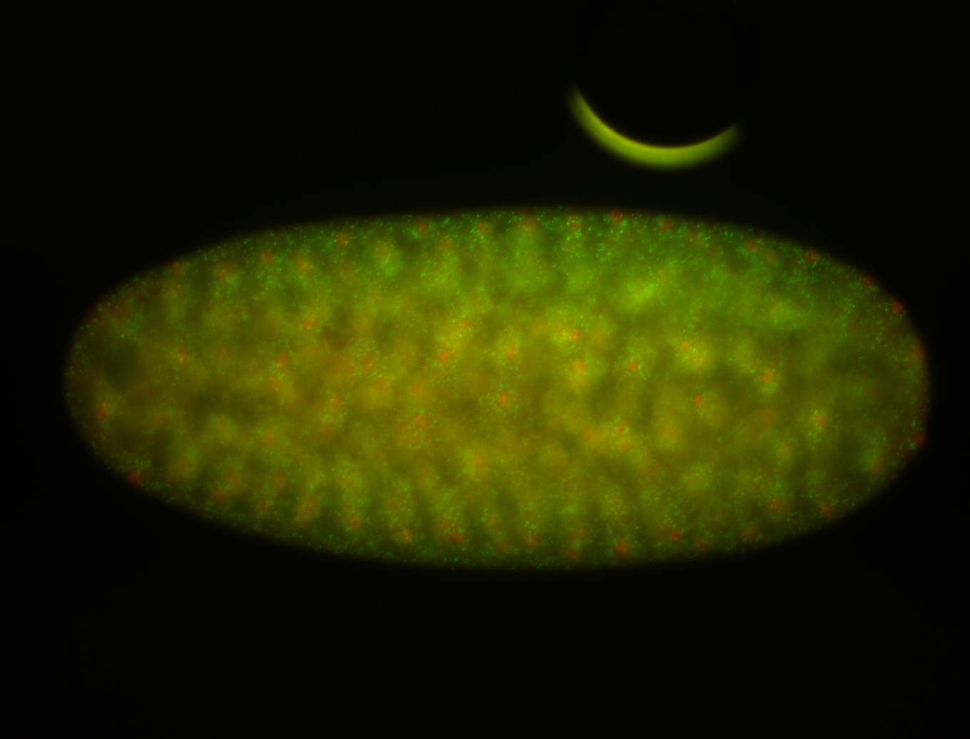
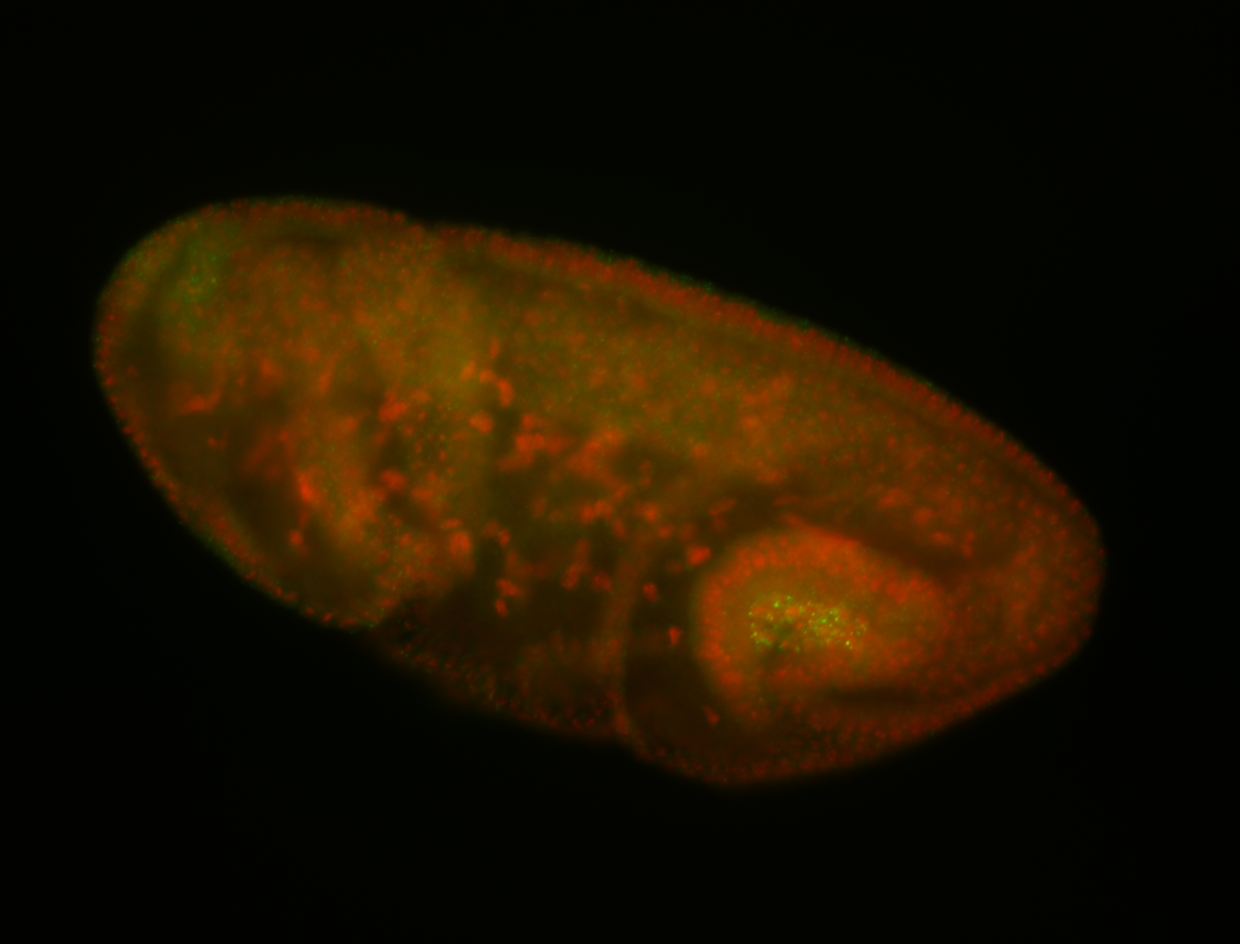 | Apical enrichment Pole cell localization Discrete apical foci Localized Expressed Degraded Subset blastoderm nuclei Apical localization Degraded partially stage 7 ant blastoderm nuclei Zygotic starting stage 7 Cytoplasmic foci Stage 6-7 Blastoderm nuclei Embryonic localization patterns Degraded completely stage 7 Zygotic Subcellular localization patterns Pole cell enrichment Maternal |
| stage8-9 | 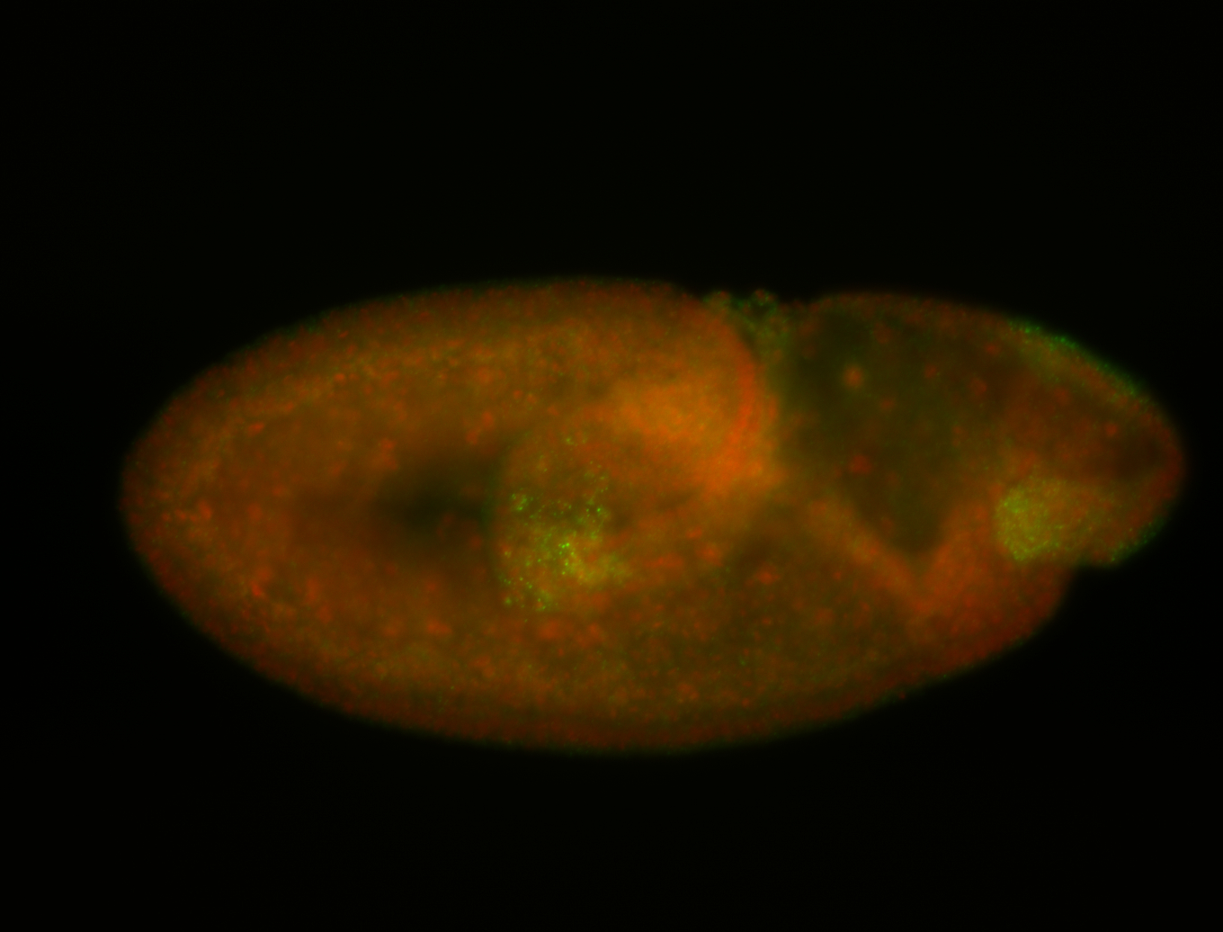 | ant blastoderm nuclei Zygotic Subcellular localization patterns Subset blastoderm nuclei Expressed Germ cell enrichment Localized Germ cell localization Embryonic localization patterns Stage 8-9 Cytoplasmic foci Blastoderm nuclei Maternal |
| Probe source: | RE24064 (Flybase link CG10360 , FlyFish insitu , BDGP insitu new) | |
| stage | images | Annotation |
| stage1-3 | 
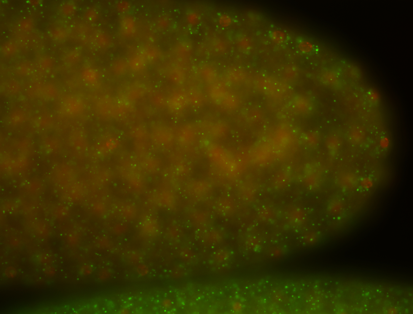
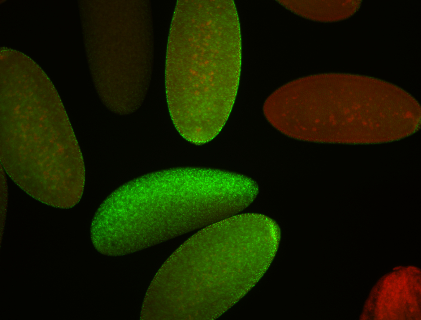
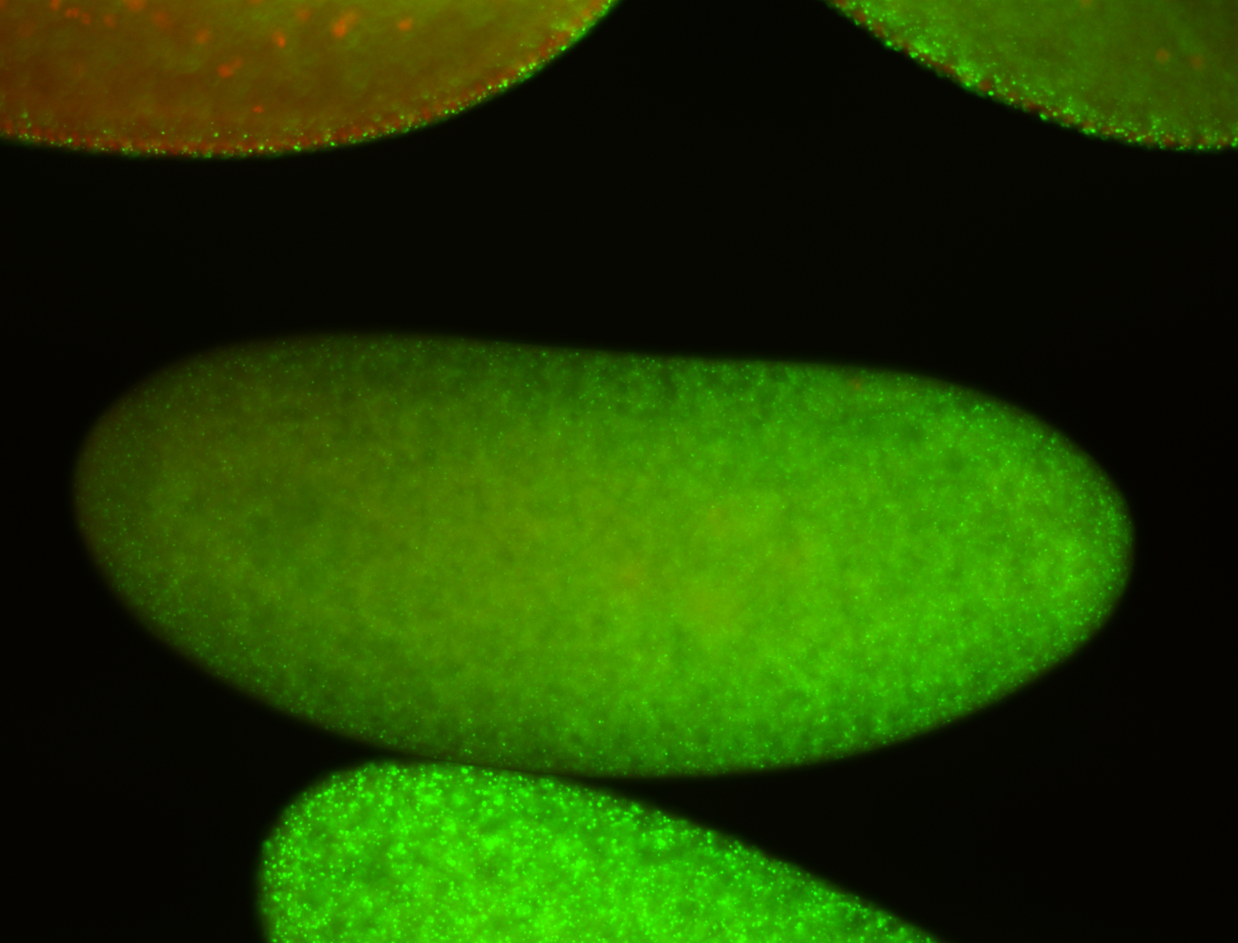
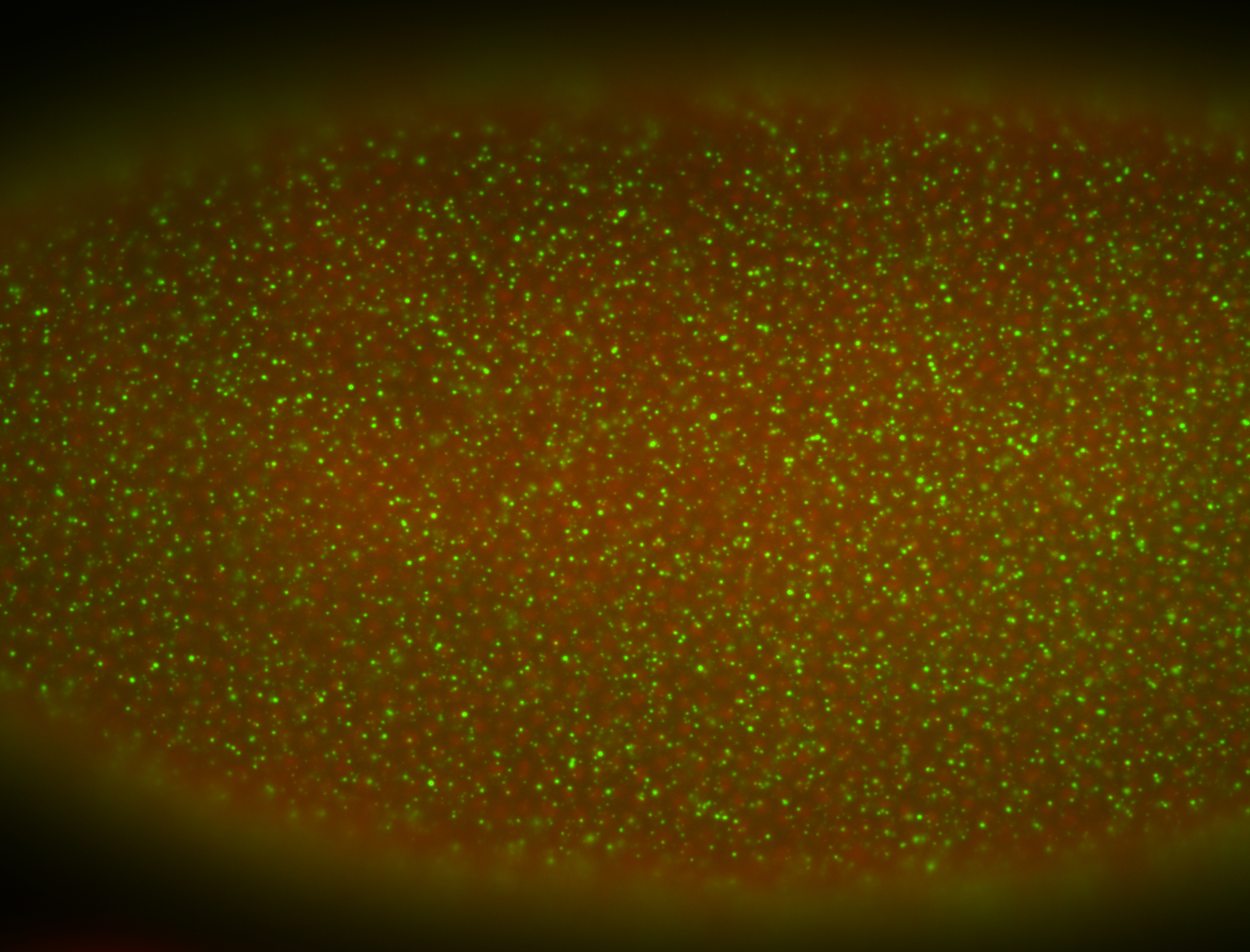

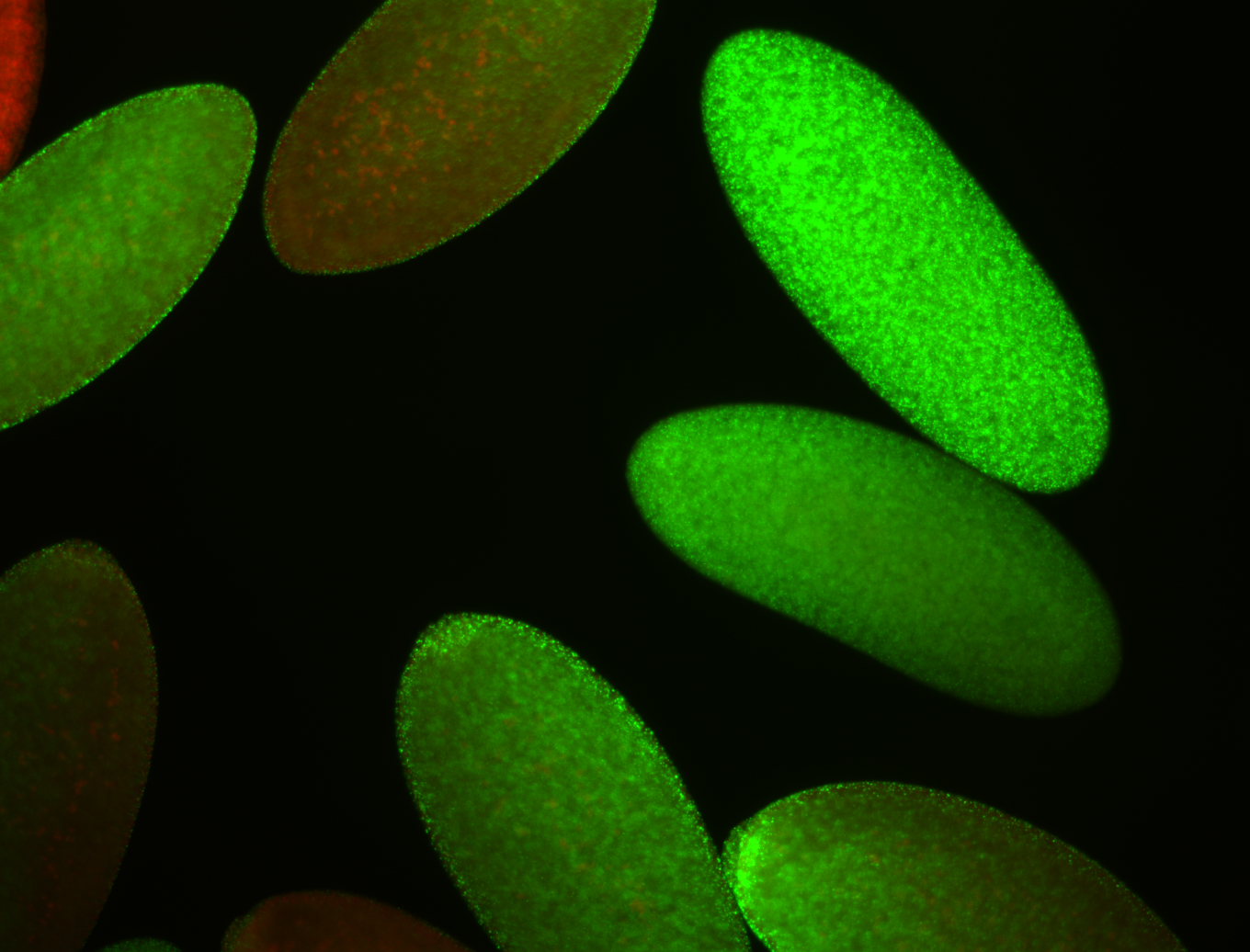
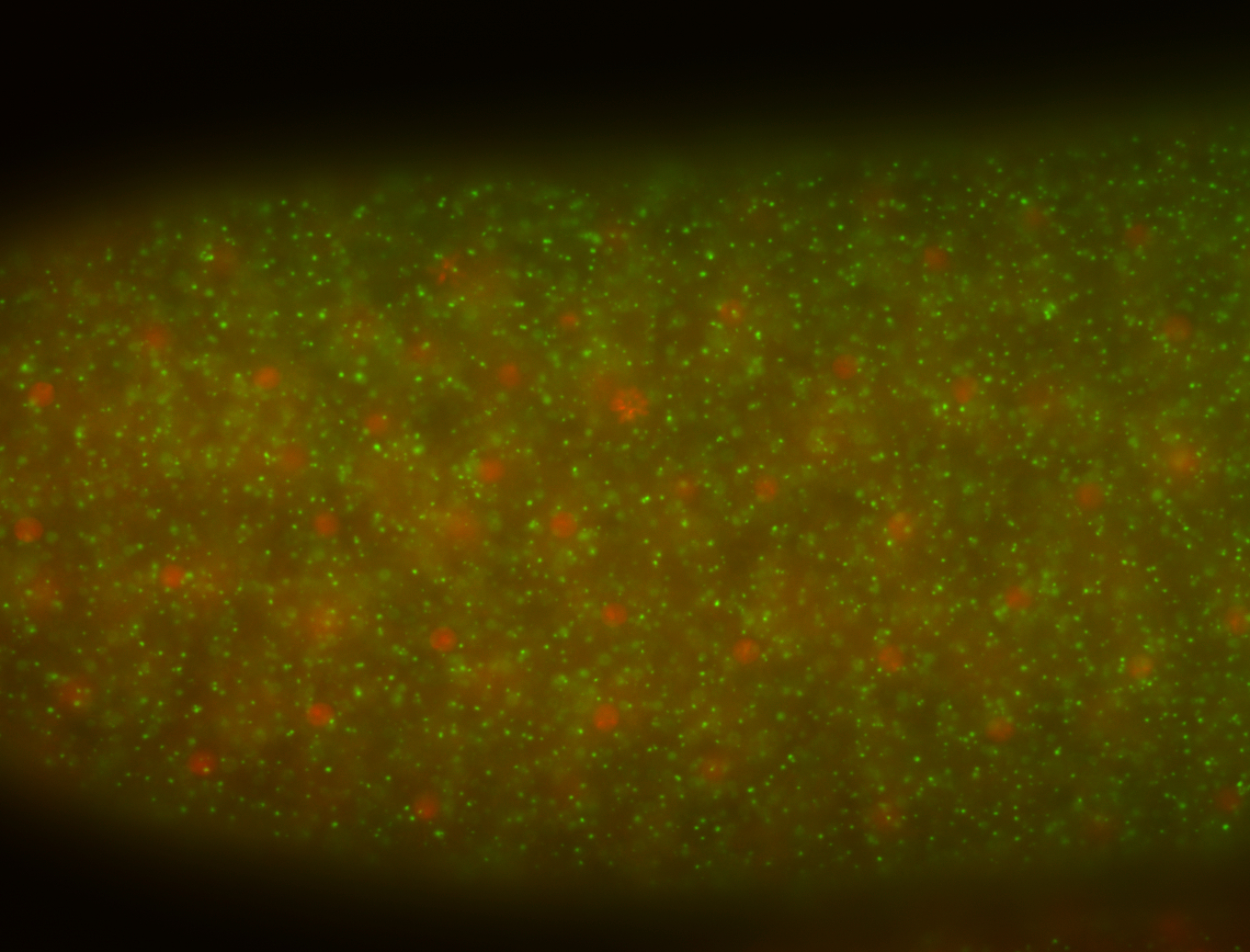
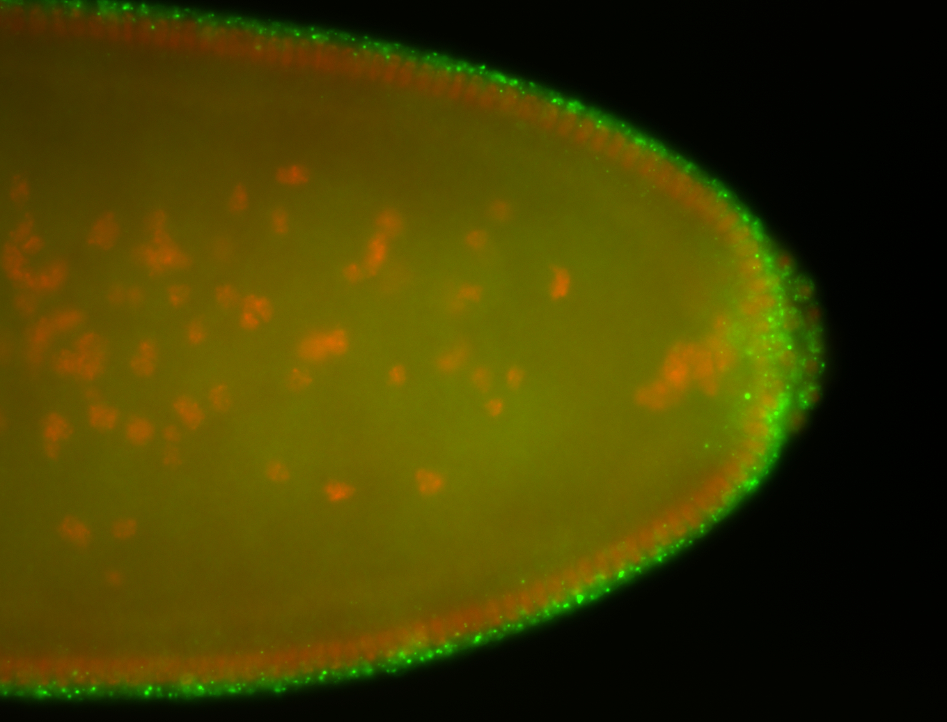
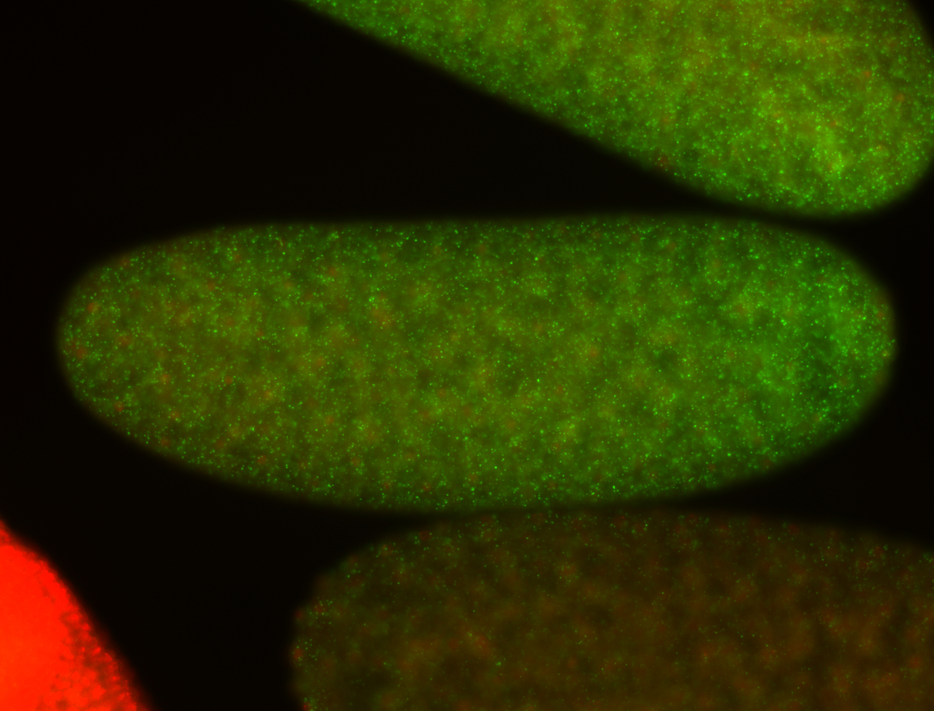
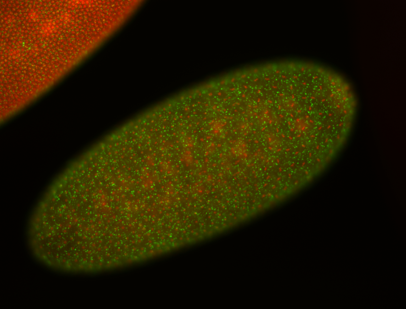
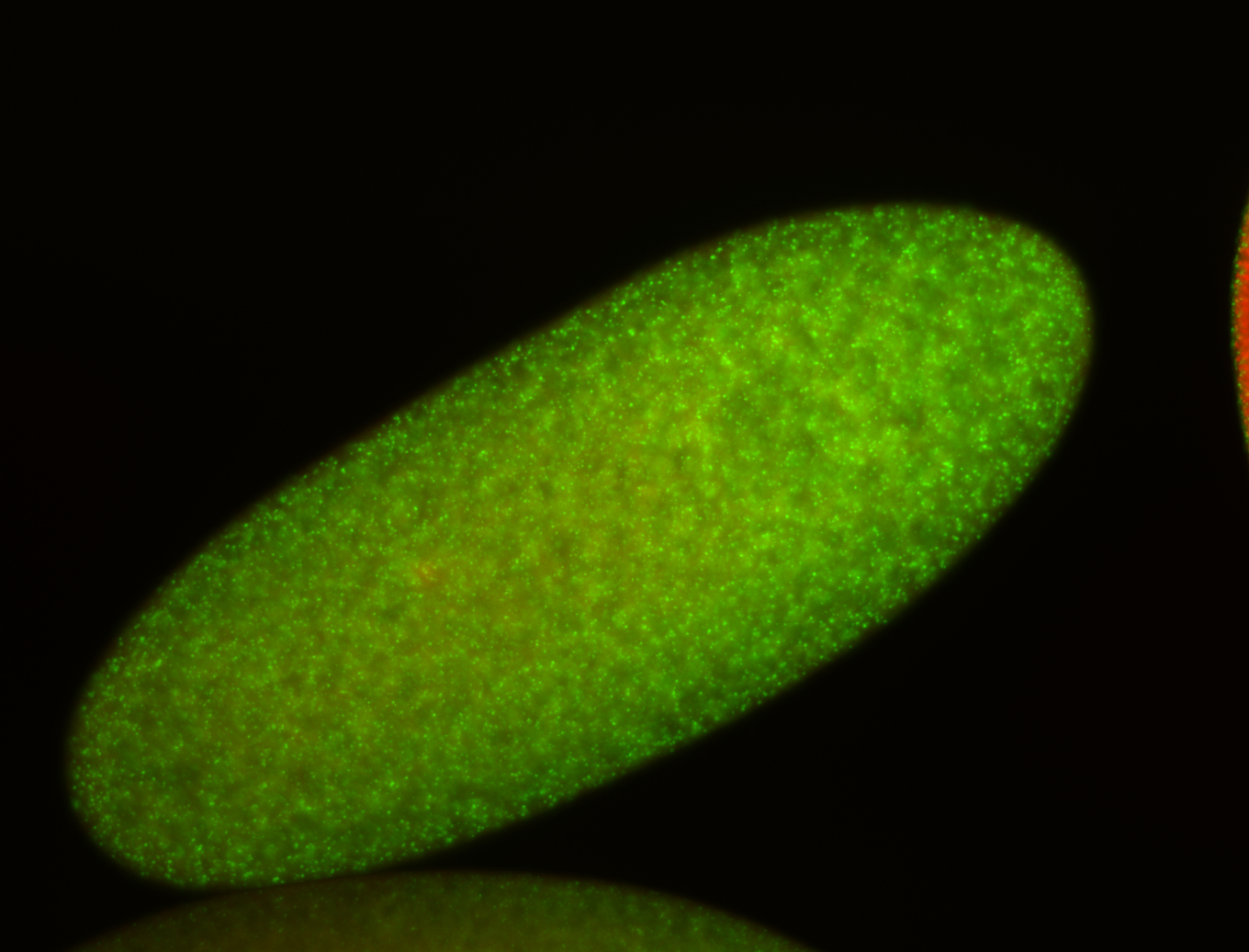
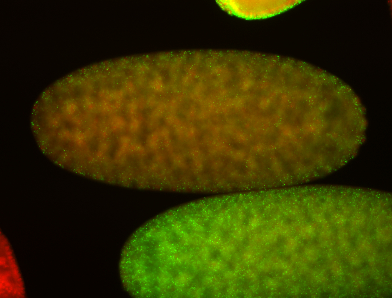


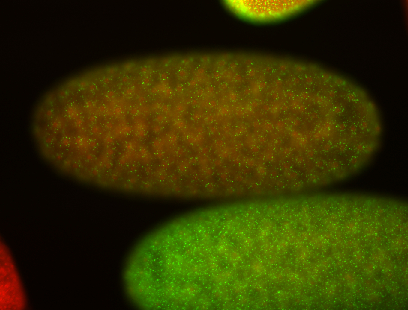 | Perinuclear localization Medium expression (21-40ms exp) Stage 1-3 Subcellular localization patterns Embryonic localization patterns Nuclei associated localization Annotated by Ronit Cytoplasmic foci Expressed Perinuclear around all nuclei Localized Maternal |
| stage4-5 | 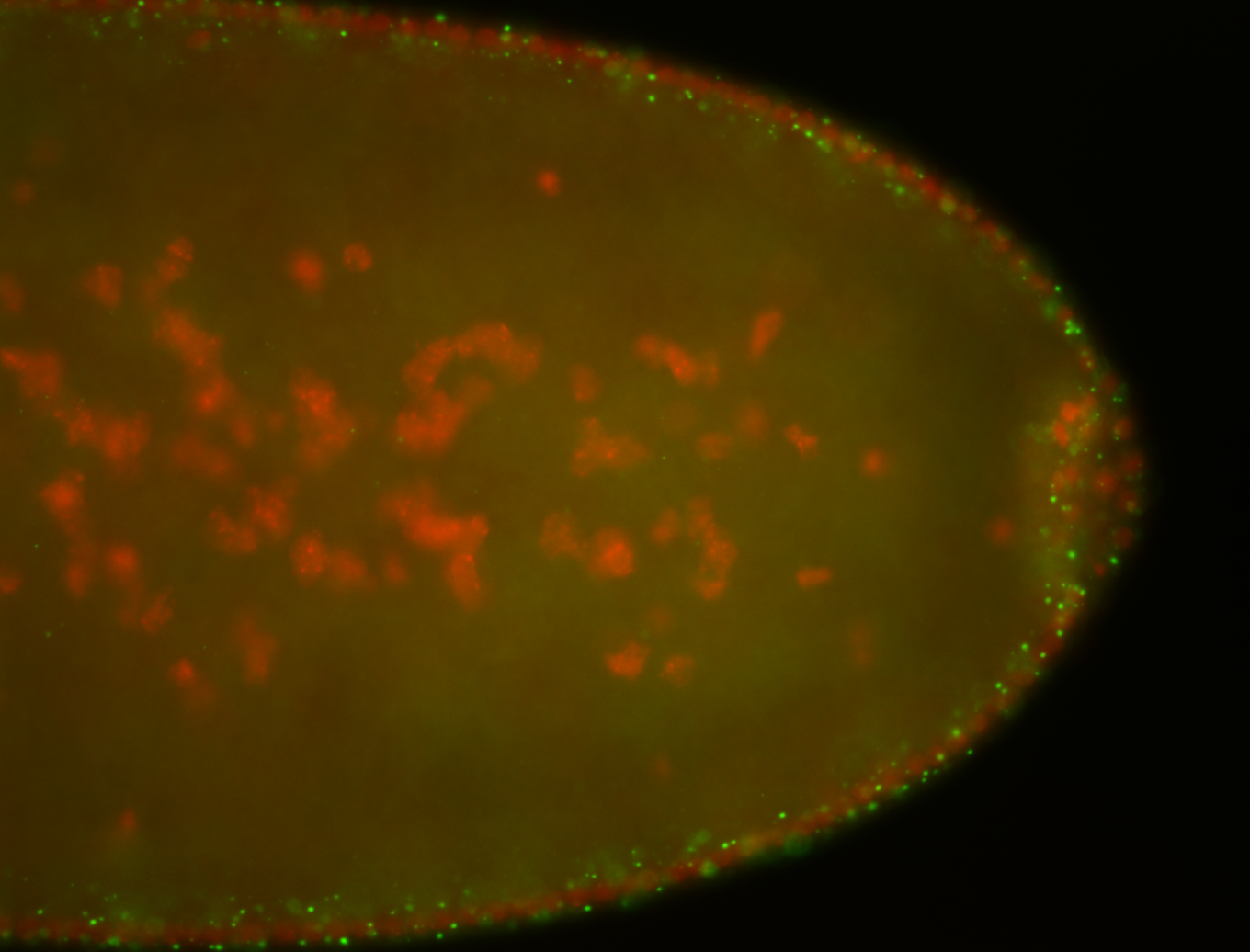
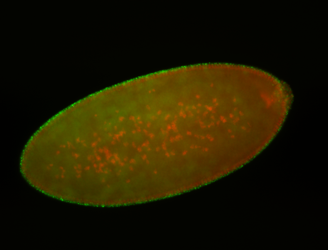
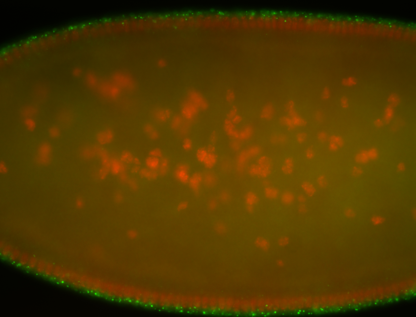
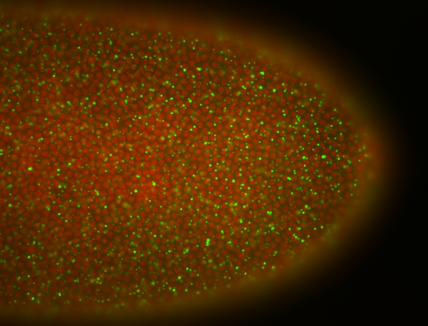
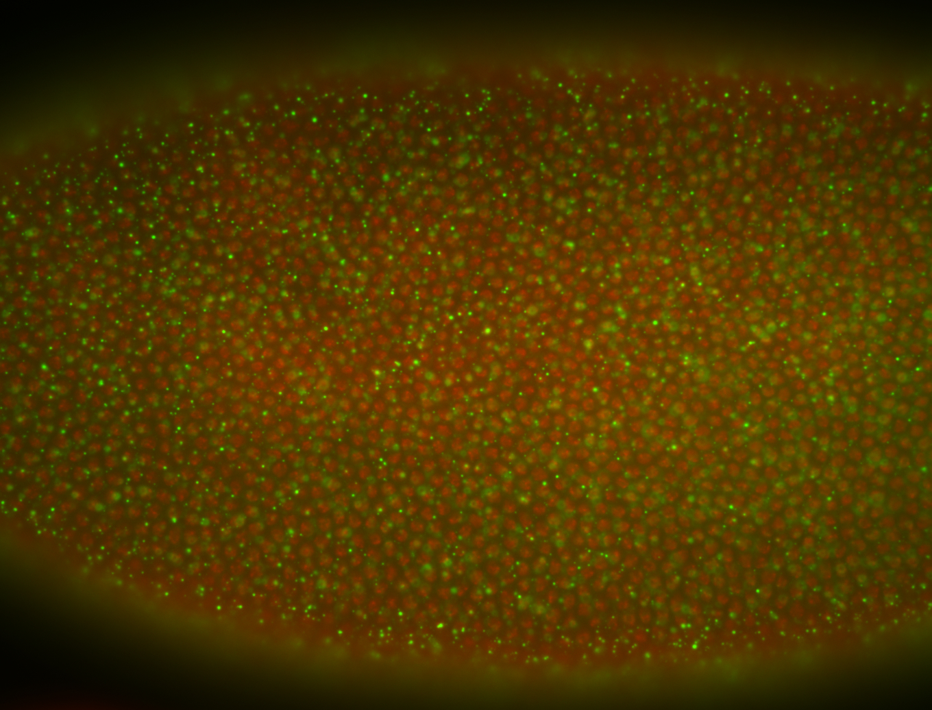
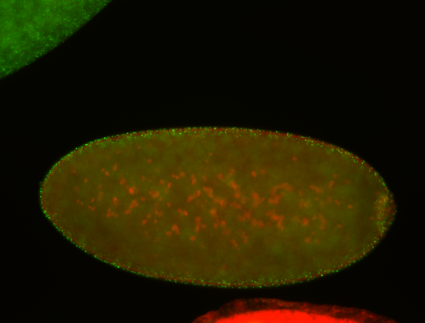
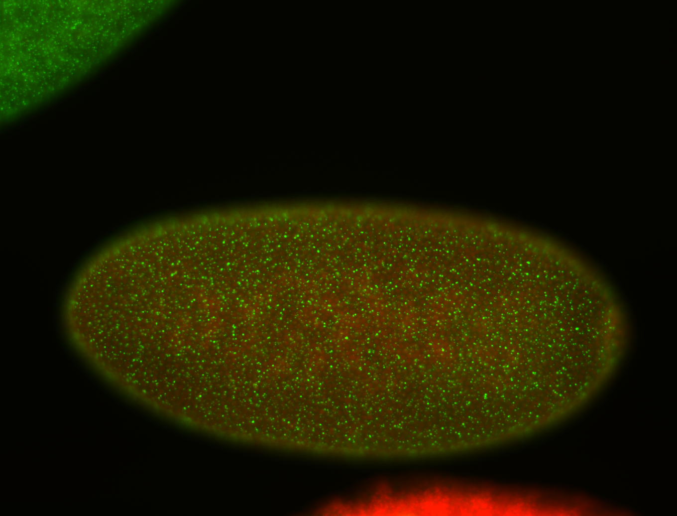

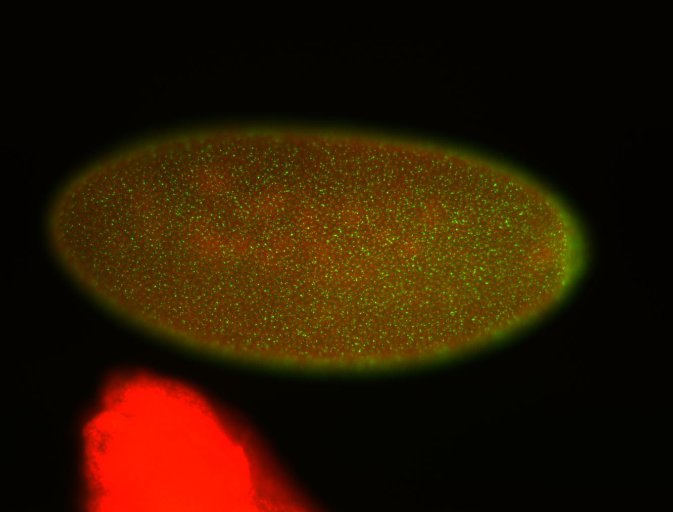
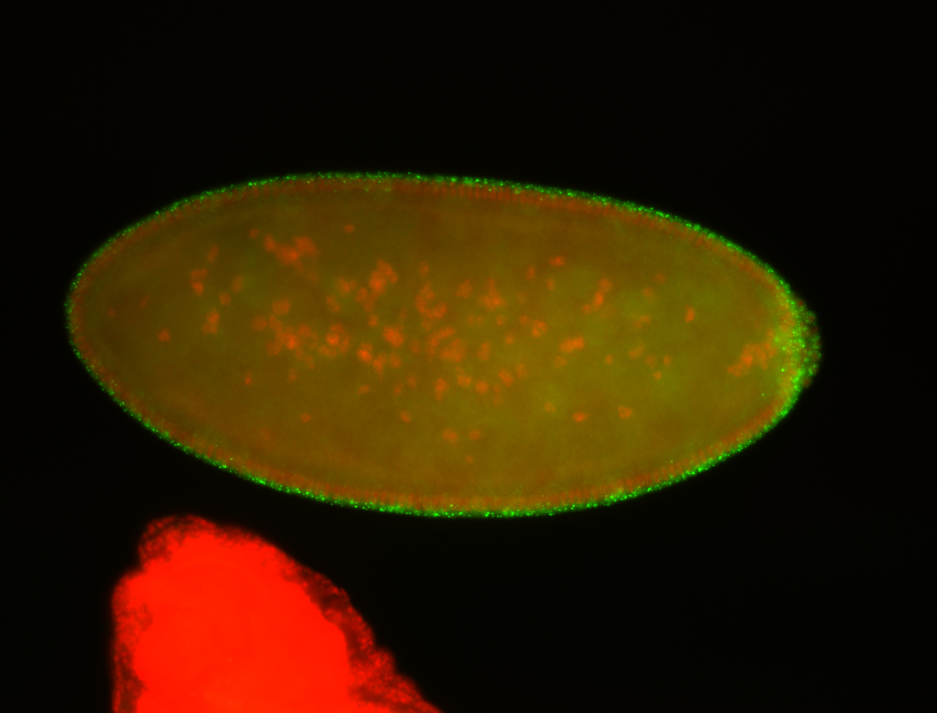
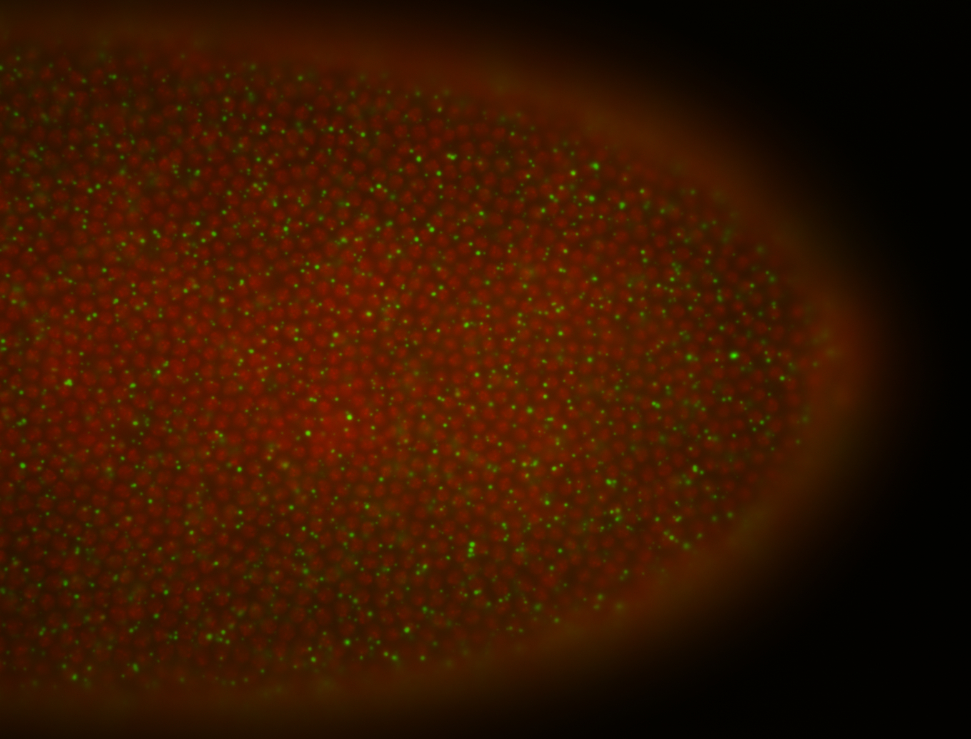 | Cytoplasmic foci post localization Embryonic localization patterns Subcellular localization patterns Medium expression (21-40ms exp) Basal exclusion Perinuclear around pole cell nuclei Perinuclear localization Localized Expressed Apical enrichment Pole cell localization Exclusionary patterns Stage 4-5 Nuclei-associated localization Discrete apical foci Apical localization Maternal |
| stage6-7 | 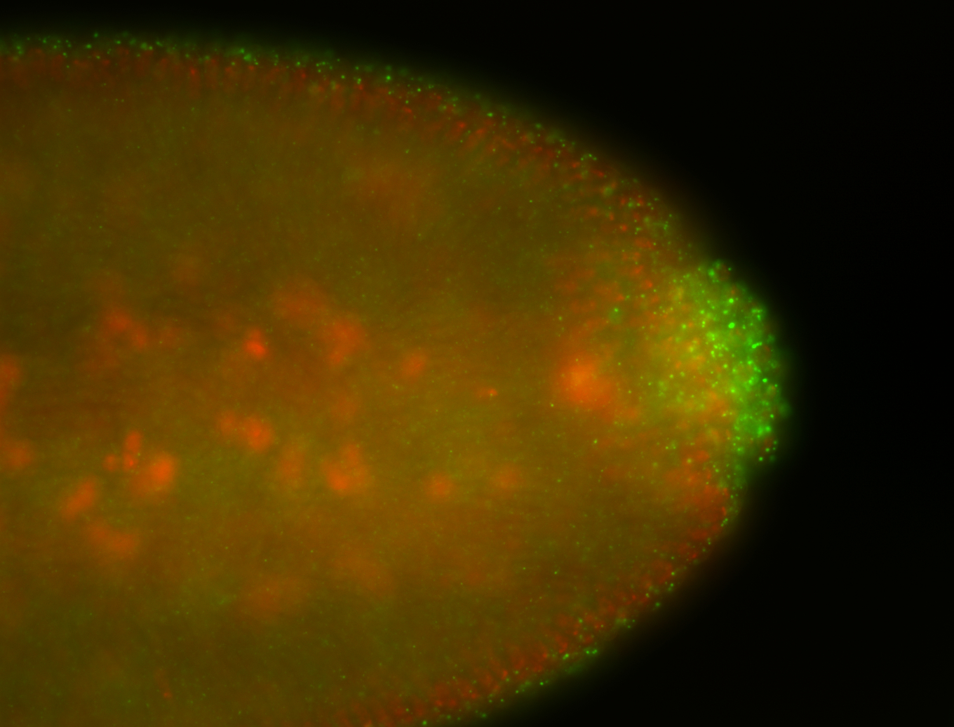
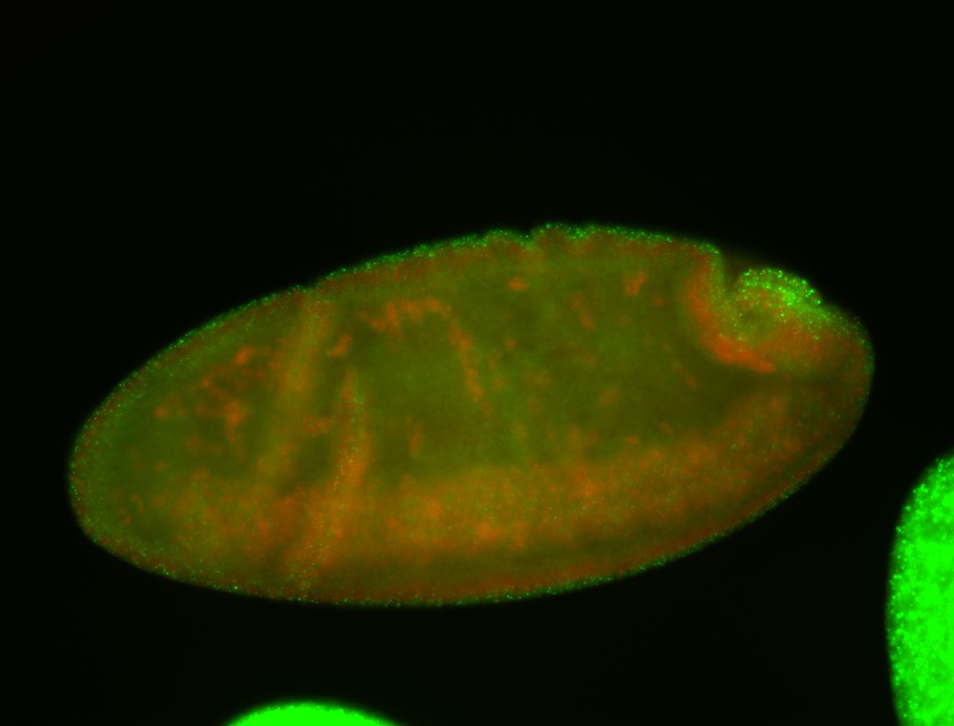
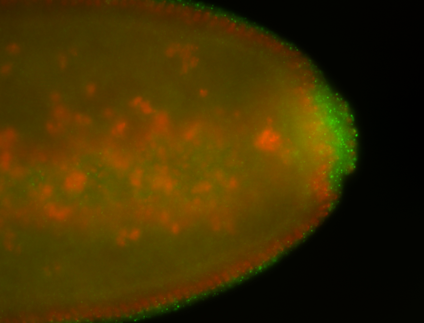
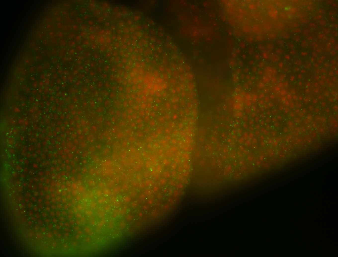
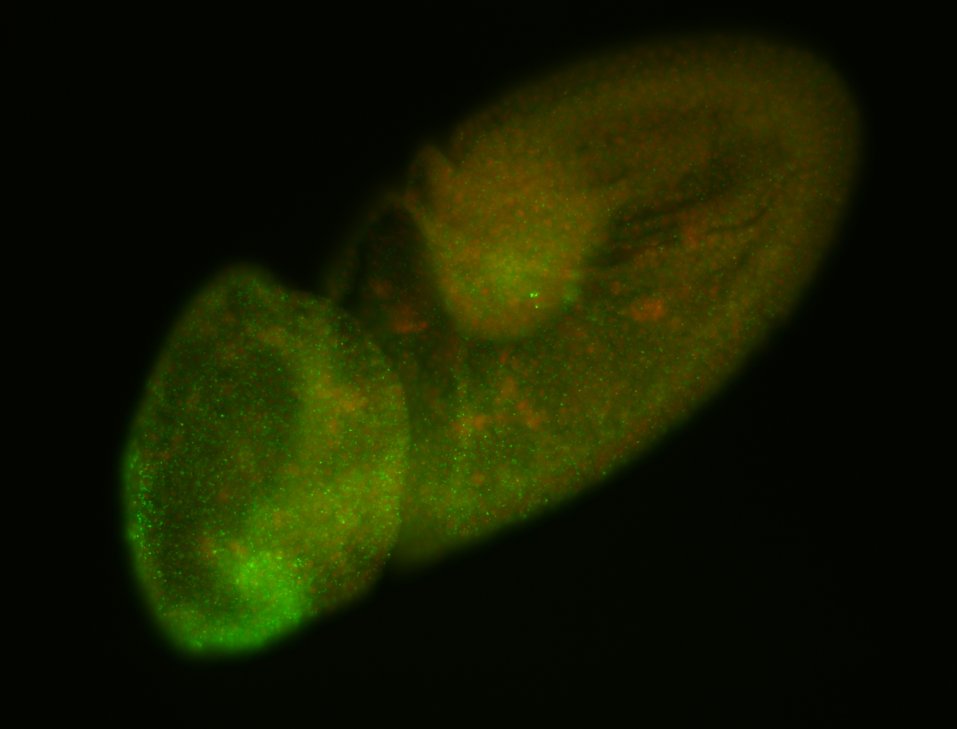
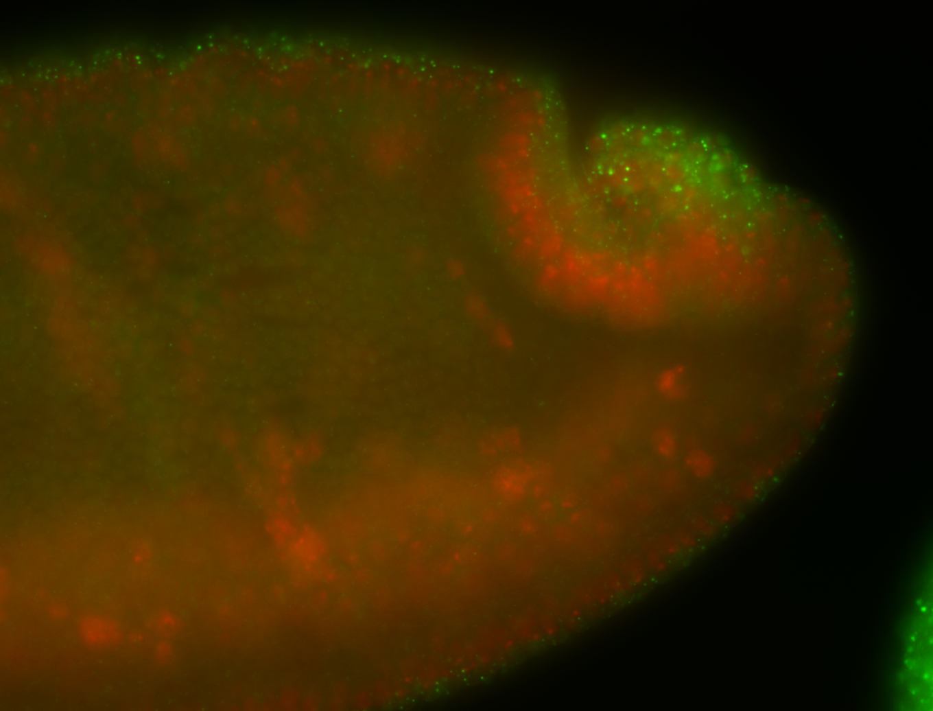
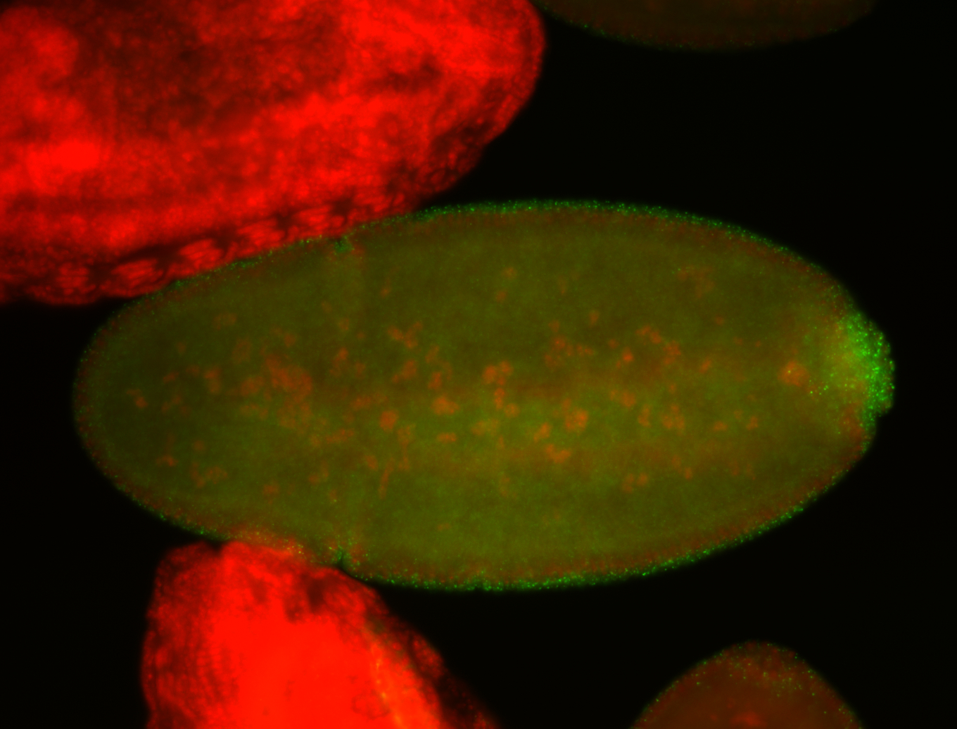
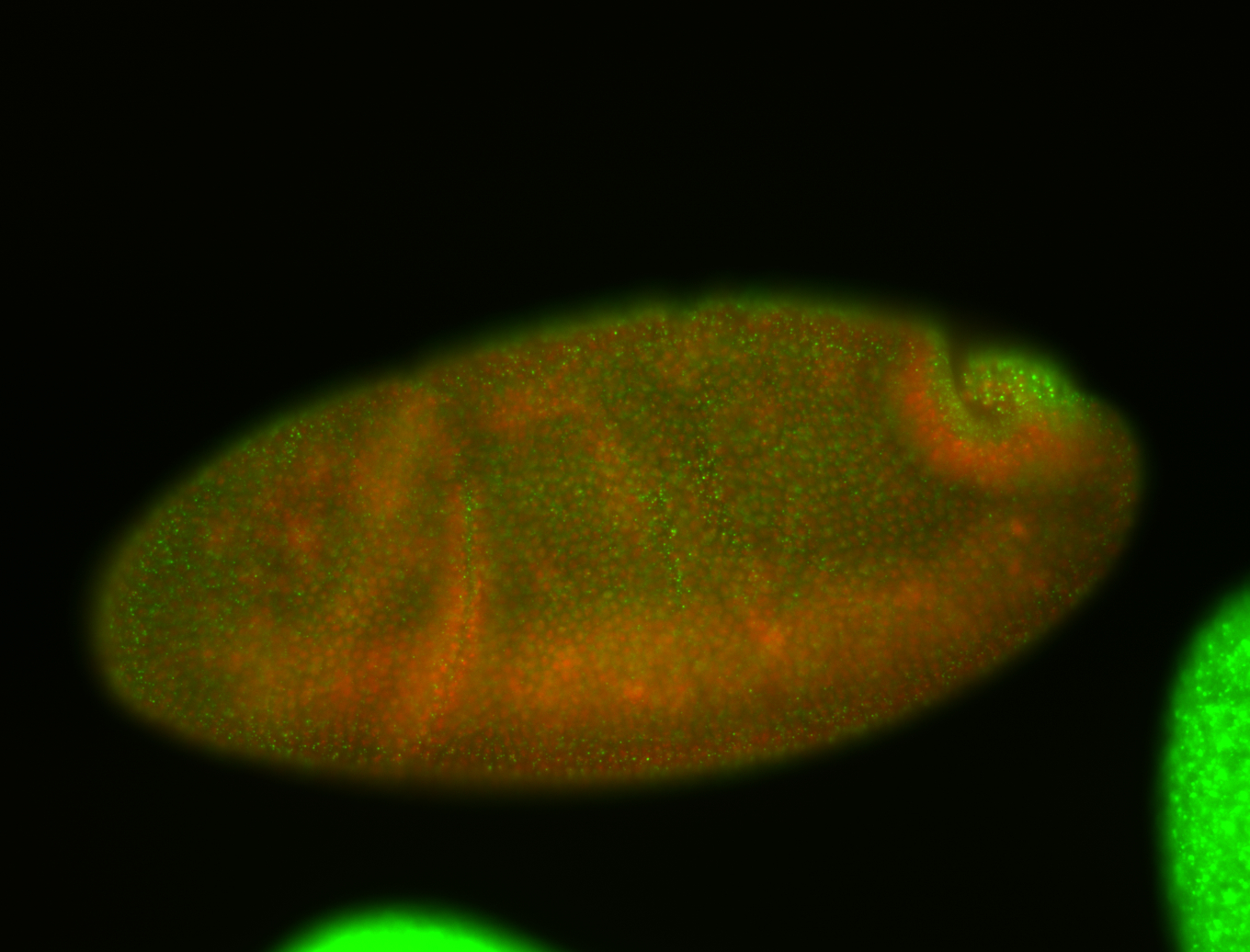
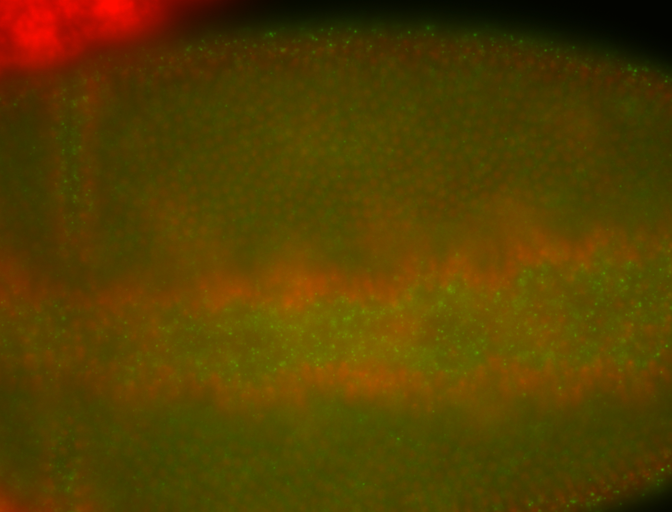
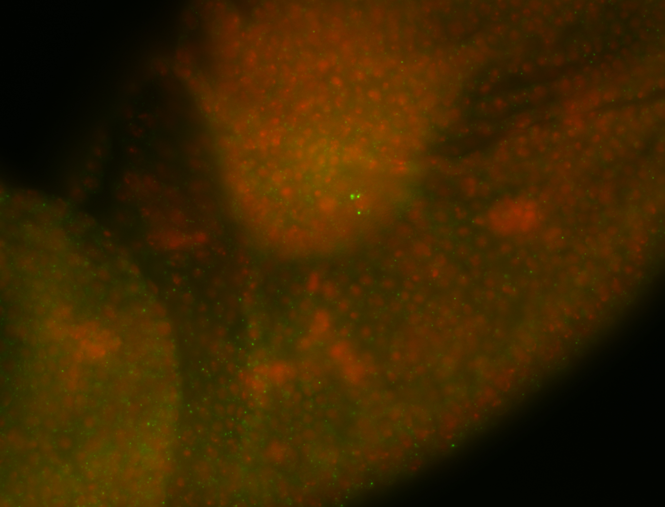 | post localization Stage 6-7 Cytoplasmic foci Blastoderm nuclei Embryonic localization patterns Zygotic Subcellular localization patterns Pole cell enrichment Medium expression (21-40ms exp) Perinuclear around pole cell nuclei Perinuclear localization Localized Expressed Pole cell localization Apical enrichment Nuclei-associated localization Discrete apical foci ant blastoderm nuclei Random blastoderm nuclei Apical localization Subset blastoderm nuclei |
| stage8-9 | 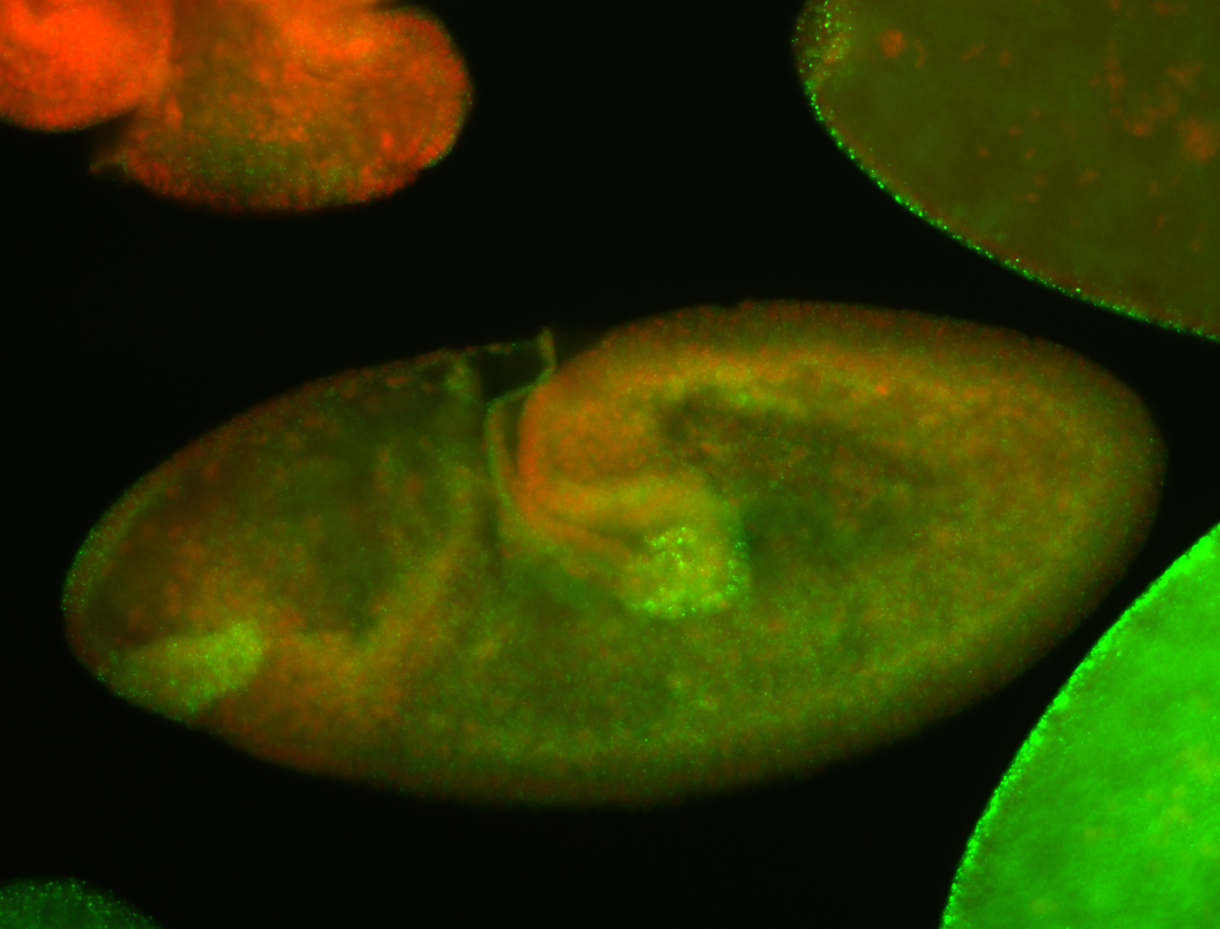
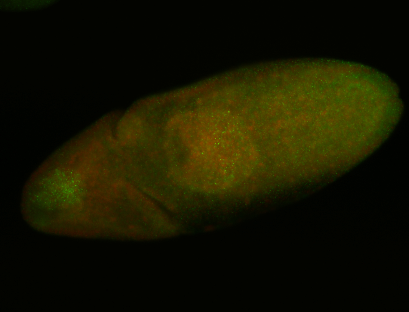

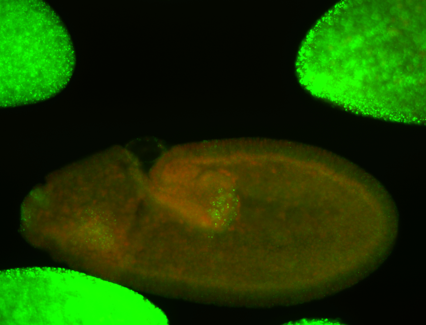
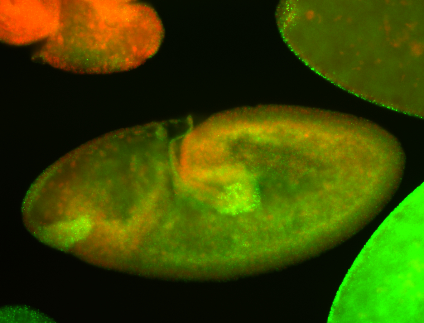 | ant blastoderm nuclei Subcellular localization patterns Zygotic Subset blastoderm nuclei Perinuclear around pole cell nuclei Perinuclear localization Weak expression (>41ms exp) Degraded Germ cell nuclei Expressed Localized Nuclei-associated localization Stage 8-9 Blastoderm nuclei Degraded partially stage 9 |
| stage10- | 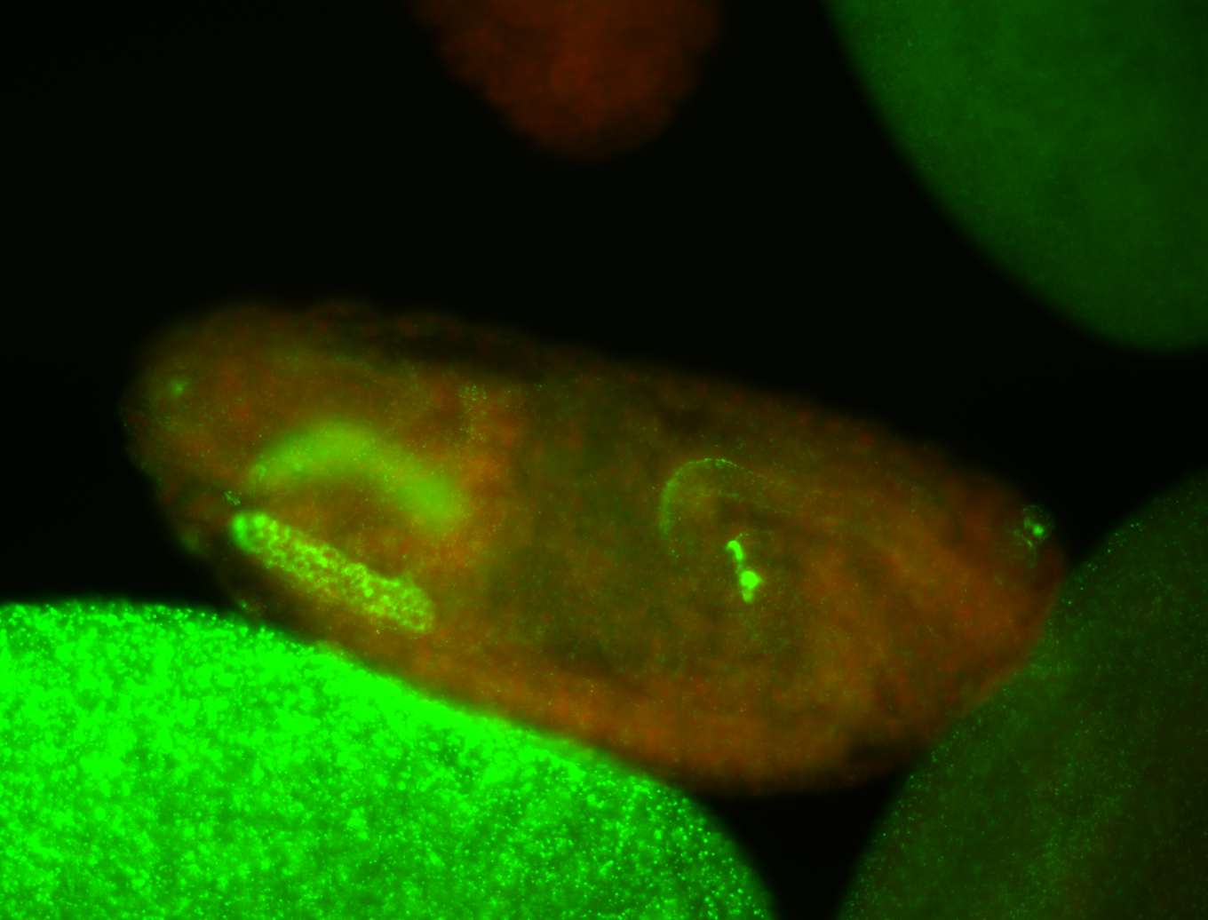
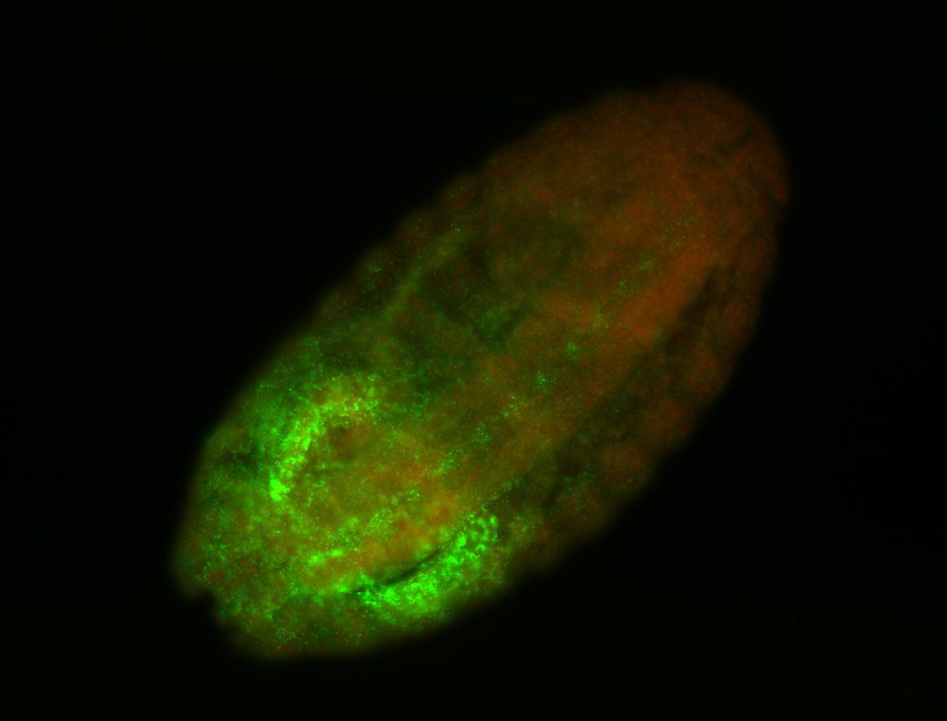

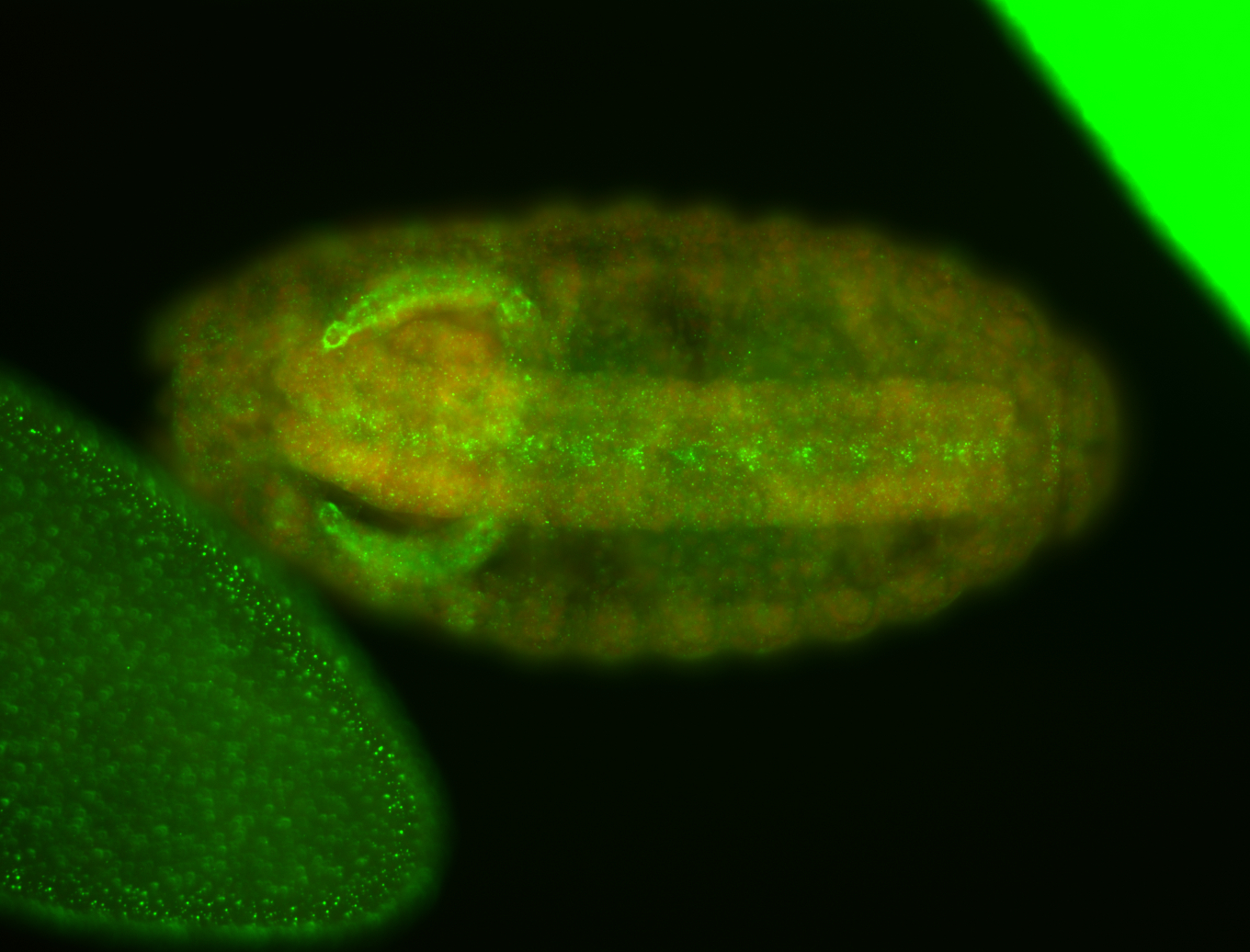 | Blastoderm nuclei Localized Hindgut Expressed Specific hindgut area or cells post spiracles Zygotic Subcellular localization patterns Salivary glands - specific region Stage 10- Salivary glands vent nerve cord |
Annotation summary for ref(2)P in insitu
Legend: RNA signal: green, DNA staining: magenta
| Probe source: | RE24064 (Flybase link CG10360 , FlyFish insitu , BDGP insitu new) | |
| stage | images | Annotation |
| stage1-3 | maternal | |
| stage4-6 | list_4-6:specific ubiquitous | |
| stage7-8 | ubiquitous pole cell | |
| stage9-10 | ubiquitous procephalic ectoderm PR trunk mesoderm PR head mesoderm PR ant endoderm PR post endoderm PR germ cell | |
| stage11-12 | ubiquitous post midgut PR ant midgut PR germ cell | |
| stage13-16 | cat_term:global_finito ubiquitous hindgut vent midline crystal cell midgut | |
| Probe source: | GH06306 (Flybase link CG10360 , FlyFish insitu , BDGP insitu new) | |
| stage | images | Annotation |
| stage1-3 | maternal | |
| stage4-6 | list_4-6:specific cellular blastoderm | |
| stage7-8 | list_7-8:specific trunk mesoderm PR post endoderm PR ant endoderm A pole cell | |
| stage9-10 | trunk mesoderm PR ant endoderm PR | |
| stage11-12 | germ cell | |
| stage13-16 | germ cell | |
| Probe source: | GH06306-dg (Flybase link CG10360 , FlyFish insitu , BDGP insitu new) | |
| No images or annotation terms | ||